
II. Physiology: Neuromuscular - Sensory
- Sensory feedback is critical to maintaining continuous fluid movement that balances various motor Neuron inputs
- Golgi Tendon Organs (Neurotendinous Spindle)
- Detect Muscle tendon tension
- Measures combination of Muscle Contraction against the resistance of opposing forces (gravity, Antagonist Muscles)
- Triggers a reduction in Muscle tension by allowing further Muscle stretch
- Muscle Spindles
- Wrap intrafusal Muscle fibers
- Small Muscle fibers primarily for stretch detection
- Intrafusal Muscle fibers are innervated by gamma Motor Neurons
- Contrast with the extrafusal Muscle fibers, the primary, major Muscle fibers of motor activity
- Extrafusal Muscle fibers are innervated by alpha Motor Neurons
- Alpha and gamma Neurons typically fire together, keeping extrafusal and intrafusal Muscles in sync
- Small Muscle fibers primarily for stretch detection
- Detect Muscle stretch (and the speed of Muscle stretch)
- Transmit stretch Sensation to the spinal cord and Cerebellum
- Increased detected Muscle stretch drives increased counter Muscle Contraction
- Rapidly increased stretch, results in stronger signaling for Muscle Contraction
- Deep Tendon Reflexes generate a rapid stretch impulse with an exaggerated Muscle Contraction
- Primary nerve endings (Type 1A, Annulospiral)
- Detect degree and rate of Muscle stretch
- Secondary nerve endings (Type 2)
- Detect degree of Muscle stretch
- Wrap intrafusal Muscle fibers
- Other sensory input
- Joint capsule and ligament receptors
III. Physiology: Neuromuscular - Motor and Muscle Contraction
-
Antagonistic Muscles are inhibited during Muscle Contraction
- Triceps Muscle is inhibited during biceps flexion
- Hamstring Muscles are inhibited during quadriceps flexion
- Striated Muscle
- Muscle Cells or Fibers are composed of hundreds of Myofibrils
- Myofibrils are divided into repeated sections of Sarcomeres
- Sarcomeres are composed of overlapping myosin filaments and actin filaments
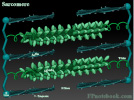
- Each Sarcomere is bordered by a Z-Disc on each end
- Actin filaments are attached to each Z-Disc
- Actin filaments do not extend completely between the Z-Discs
- A gap (H Band) divides the actin filaments facing one another
- Myosin filaments extend between the Z-Discs
- Myosin is not attached to the Z-Discs, leaving a space around each Z-Disc (I band)
- Myosin overlaps the actin filaments and appears as an A band
- Muscle Cells have additional specialized components
- Sarcoplasmic Reticulum (see below)
- Large number of mitochondria for ATP supply to fuel Muscle Contractions
- Motor units
- Motor Units represent a peripheral motor Neuron's branches and the Muscle Cells they innervate
- Neuromuscular Junction
- Neuron branches terminate at motor end plates (concentrated at center of Muscle Cells)
- When Motor Neurons fire, they typically release Acetylcholine at the motor endplate
- Strength and duration of Muscle Contraction is related to repetitive Muscle fiber discharges
- Acetylcholine acts at cell membrane of Transverse Tubule (T-Tubule)
- T-Tubules are deep invaginations into the Muscle Cell
- Acetylcholine (and other stimuli) trigger changes in Muscle cell Membrane Potential
- Action Potential propagates along the cell membrane from Muscle Cell center to its periphery
- Acetylcholinesterase within Neuromuscular Junction inactivates Acetylcholine
- Prevents prolonged Acetylcholine activity at the motor end plate
- Sarcoplasmic Reticulum
- Sarcoplasmic Reticulum is a specialized endoplasmic reticulum, positioned between the T-Tubules
- Sarcoplasmic Reticulum contains large concentrations of Calcium
- When triggered by Action Potential, Calcium is released into the Muscle Cell
- In cardiac Muscle Cells, interstitial fluid Calcium also influxes into the cell
- Muscle Contraction
- Increased Muscle Cell Calcium triggers Actin and Myosin to increase their overlap
- Muscle Relaxation

IV. References
- Goldberg (2014) Clinical Physiology, Medmaster, Miami, p. 90-5