
II. Exam: Telemedicine
- See Telemedicine
- Both knees should be exposed (e.g. shorts)
- Evaluate patients gait from front and side (see standing exam below)
- Perform knee general exam as below (observation, self-palpated tenderness and range of motion)
- Perform specific knee tests as able (see below)
- Consider Knee XRay or other imaging indications
- Ottawa Knee Rule (if Knee Pain after Trauma)
- Inability to actively extend the knee (e.g. Patella injury, quadriceps Tendon Injury)
III. Exam: General (compare with less affected knee)
- Observation
- Obvious injury findings
- Deformity
- Ecchymosis
- Overlying skin changes
- Erythema
- Open wound (e.g. Open Joint Injury, Open Fracture)
- Skin Tenting
- Previous surgical scars
- Knee Effusion or swelling with obscured landmarks
- Quadriceps Muscle atrophy
- Evaluate Vastus Medialis Obliquus specifically
- Atrophy often on side of Ligamentous Injury
- Obvious injury findings
- Tenderness to Palpation
- Anterior (see Patella below)
- Patella (Fracture, Contusion, Prepatellar Bursitis)
- Tibial tubercle (avulsion Fracture, Patellofemoral Syndrome, Osgood-Schlatter)
- Patellar tendon (Patellar Tendonitis, Sinding-Larsen-Johansson Syndrome)
- Quadriceps tendon (e.g. Quadriceps Tendonitis)
- Medial
- Medial Meniscus Tear (joint line tenderness)
- Osteoarthritis
- Medial plica (tender Nodule at joint line)
- Pes Anserine Bursitis (3 cm inferior to anteromedial joint line)
- Medial collateral ligament tear
- Medial Tibial Plateau Fracture
- Osteochondral or subchondral insufficiency Fracture
- Lateral
- Iliotibial Band Syndrome (proximal tibial tenderness at IT Band insertion, Gerdy Tubercle)
- Lateral Meniscal Tear
- Lateral collateral ligament tear
- Lateral Tibial Plateau Fracture
- Osteochondral or subchondral insufficiency Fracture
- Proximal fibular Fracture (e.g. Maisonneuve Fracture)
- Posterior
- Popliteal Cyst (Baker's Cyst)
- Gastrocnemius Tear (typically medial gastrocnemius tendon)
- Posterior Cruciate Ligament Tear
- Tear of posterior aspect of meniscus
- Hamstring injury or tenderness
- Semimembranosus Tendonitis (posteromedial)
- Semitendinosus Tendonitis (posterolateral)
- Anterior (see Patella below)
-
Knee Range of Motion
- Note knee resting position
- Normal range of motion
- Flexion: 135 degrees
- Extension: 0 to -10 degrees (above horizontal plane)
- Significant effusion or Monoarthritis may limit knee extension to <25 degrees from a resting flexed position
IV. Exam: Patella
- Patella deformity or focal tenderness
- Quadriceps tendon insertion tenderness
-
Patellar tendon insertion tenderness (inferior pole Patella)
- Patellar Tendonitis (Jumper's Knee)
- Sinding-Larsen-Johansson Syndrome (early teens)
- Tenderness to the medial or lateral aspect of the Patellar tendon (but not immediately over the tendon)
- Infrapatellar fat pad (Hoffa's fat pad)
- Tibial Tuberosity Tenderness
- Osgood-Schlatter Disease (teens)
- Patellofemoral Syndrome
- Tibial tubercle avulsion (with Trauma History)
-
Patellofemoral Syndrome
- Quadriceps Femoris Muscle Angle (Q Angle)
-
Patella tracking with quadriceps contraction
- Evaluate for smoothness of motion and crepitation
- Patellar Apprehension Test
V. Exam: Ligaments
- Anterior Cruciate Ligament (ACL) Stability Tests in ACL Tear
- Lever Test (most sensitive)
- Lachman Test (second most sensitive)
- Knee Anterior Drawer Test
- Pivot Shift Test (MacIntosh Test)
- Posterior Cruciate Ligament (PCL) Tests in PCL Tear
- Collateral ligament evaluation
- Knee Valgus Stress Test (Medial collateral ligament tear)
- Knee Varus Stress Test (Lateral collateral ligament tear)
VI. Exam: Meniscus
- See Meniscus Injury
- McMurray's Test
- Apley's Compression Test and Apley's Distraction Test
- Knee Bounce Test
- Thessaly Test
- Inability to fully extend knee may suggest "bucket-handle" meniscal tear
- Joint line tenderness is 76% sensitive for meniscal tear, but not specific
VII. Exam: Neurovascular
- See Sensory Exam
- Lower extremity Motor Exam
- See Motor Exam
- Femoral Nerve (L1-L4)
- Hip flexion against resistance (Iliopsoas Muscle)
- Sciatic Nerve (L5-S2)
- Deep Peroneal Nerve (L4-L5): Foot Drop
- Dorsiflexion against resistance (tibialis anterior Muscle)
- Tibial Nerve (S1-S2)
-
Deep Tendon Reflexes
- Patellar Reflex (L2-4)
- Achilles Reflex (S1)
- Distal pulses
- Miscellaneous
- Compartment Syndrome findings (significant limb circumferential swelling, pain out of proportion)
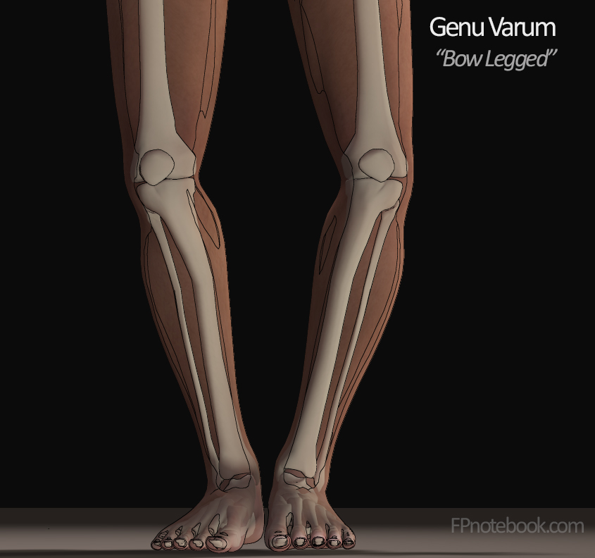
VIII. Exam: Standing Evaluation

IX. Imaging
- See Knee Imaging
- See Knee XRay
- See Knee XRay Indications in Acute Injury (e.g. Ottawa Knee Rules)
- See Knee Ultrasound
X. References
- Bach (1997) Physician Sportsmed, 25(5): 39-50
- Budoff (1997, April) Consultant, 919-30
- Budoff (1997, Feb) Consultant, 295-304
- Hoppenfeld (1976) Physical Exam, Prentice-Hall
- Bunt (2018) Am Fam Physician 98(9): 576-85 [PubMed]
- Calmbach (2003) Am Fam Physician 68(5):907-12 [PubMed]
- Pendergraph (2026) Am Fam Physician 114(1): 49-62 [PubMed]
- Rothenberg (1993) Postgrad Med, 93(3): 75-86 [PubMed]
- Smith (1995) Am Fam Physician 51(3):615-21 [PubMed]
- Solomon (2001) JAMA 286:1610-20 [PubMed]
- Yedlinsky (2021) Am Fam Physician 103(3):147-54 [PubMed]