
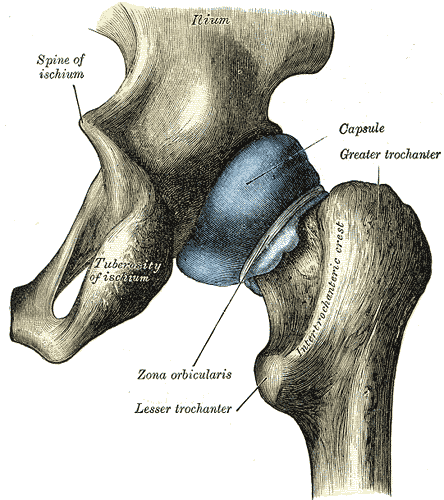
II. Anatomy
- See Hip Anatomy
- Images
 Lewis (1918) Gray's Anatomy 20th ed (in public domain at Yahoo or BartleBy)
III. Precautions: Findings most suggestive of hip intra-articular cause
- Pain on external and internal hip rotation
- Pain on hip axial loading (force applied at foot or knee towards hip)
IV. Exam: Telemedicine
- See Telemedicine
- Patient stands facing camera with feet at Shoulder width apart
- Patient places both hands with palms against the iliac crests
- Examiner compares the level of hands for symmetry (e.g. Leg Length Discrepancy)
- Examiner compares the anterior/posterior placement of hands for symmetry (pelvic rotation)
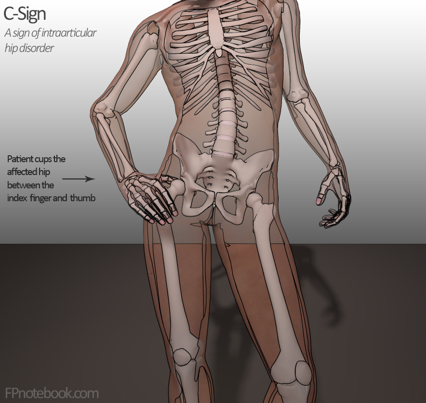
- Observe for C Sign, Cupping the painful, anterolateral hip (intraarticular hip disorder)
- Patient self-palpates regions for tenderness
- Anterior superior iliac spine
- Anterior inferior iliac spine
- Greater trochanter
- Evaluate Hip Range of Motion
- Perform hip specific testing as below
- Leg Neurologic Exam
V. Exam: External to hip
- Critical to evaluate for referred pain
- Low Back Exam (e.g. radicular pain)
- Lower extremity Neurologic Exam (sensory and motor function)
- Evaluate femoral and Pedal Pulses
- Exam of Abdomen and Pelvis
- Appendicitis or Diverticulitis may present with Hip Pain
- Other common causes of pain referred to the hip
- Knee Exam
- Greater trochanter tenderness to palpation
VI. Exam: Hip and Groin
- Observe for groin Ecchymosis (avulsion, Muscle tear, abdominal wall Hematoma)
- Observe for bulge in the abdominal and inguinal region (Hernia)
- Also palpate the Superficial Inguinal Ring with valsalva or cough
- Examine in frog-leg position
- Palpate the lower Abdomen and pupic symphysis
- Palpate adductor insertions (pubic tubercle, medial inferior pubic ramus)
- Palpate abdominal Muscles
- Palpate anterior superior iliac spine (ASIS, sartorius and tensor fasciae latae insertion)
- Palpate anterior inferior iliac spine (AIIS, rectus femoris insertion)
- Palpate anterior hip
- Palpate greater trochanter
VII. Exam: Observation
- Resting position of the hip
- Hip deformity or swelling
- Overlying skin changes
- Hip Asymmetry
VIII. Exam: Hip Range of Motion
- See Hip Range of Motion for normal findings
- Perform active range of motion and passive range of motion
- Pain on even slight range of motion should suggest intrinsic hip pathology
- Septic Arthritis of the hip should be on the differential
- See causes of inability to bear weight below
IX. Exam: Observation of Mobility and Gait
- See Abnormal Gait
- Observe sitting, standing and Transferring
- Observe while standing
- Look for C Sign (Cupping the painful, anterolateral hip)
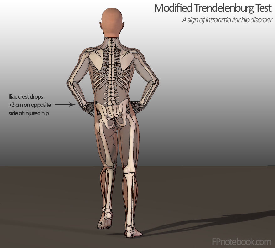
- Modified Trandelenburg Test (Single leg stance phase)
- Look for C Sign (Cupping the painful, anterolateral hip)
- Observe for limp or altered gait
- See Gait
- Trandelenburg Gait (hip adductor weakness)
- Waddling Gait
- Antalgic Gait
- Pelvic Rotational Wink
- Observe for inability to bear weight
- Hip Stress Fracture
- Hip Septic Arthritis
- Avascular Necrosis of the Hip
- Femoral lesion (e.g. malignancy)
- Observe for inability to climb onto exam table
- Decreased Hip Joint flexibility
- Iliopsoas Muscle or quadriceps Muscle Weakness
- Observe Leg Neurologic Exam
X. Exam: Specific Tests - General
-
FABER Test
- Flexion ABduction External Rotation
- Also known as Patrick's Test or Figure of Four Test
-
FADIR Test
- Flexion ADduction Internal Rotation
-
Hip Adduction Test
- Also includes Single Hip Adductor Test, Bilateral Hip Adductor Test
- Ober Test
-
Log Roll Test (Freiberg Test)
- Passive Supine Hip Rotation (with hip and knee extended)
- Stinchfield Test
XI. Exam: Specific Tests in Posterior Hip Pain (e.g. Gluteal Tendinopathy)
-
Modified Trandelenburg Test
- Standing on one leg and observe for pelvic drop on the side of the lifted leg
- Positive in Gluteal Tendinopathy or hip pathology (e.g. Hip Labral Tear, LGP, SCFE, Transient Synovitis)
-
Resisted External Derotation Test
- Lateral Hip Pain with hip extension and external rotation against resistance
- Positive in Gluteal Tendinopathy
-
Seated Piriformis Stretch Test
- Pain on ischial palpation and passive hip internal rotation
- Positive in Deep Gluteal Syndrome including Piriformis Syndrome
-
Long-Stride Walking Test
- Long step onto unaffected foot provokes Posterior Hip Pain and Sciatica in the affected hip
- Positive in Ischiofemoral Impingement Syndrome