
II. Anatomy: Groin
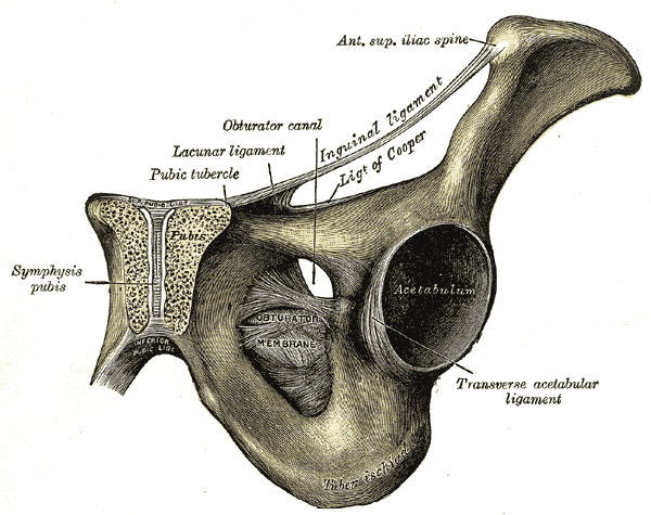
- Groin is centered around the pubic rami and pubi symphysis
- Forces generated with kicking, rotation or lateral movement
- Pubic tubercle bears a majority of the torque forces in the region
- Attaches hip adductors
- Attaches Lower abdominal Muscles
- Tendon and Ligament Insertions
- Superior
- Rectus Abdominis insertion (at superior pubic ramus, stabilizes abdominal wall)
- Medial
- Lateral
- Inguinal Ligament
- Inferior
- Adductor longus Muscle insertion (at pubis adjacent to Pubic Symphysis, stabilizes Pelvis)
- Superior
-
Muscles
- Hip Adductors
- Hip Abduction
- Gluteus Medius
- Gluteus Minimus
- Tensor fasciae latae Muscle
- Hip Flexors
- Hip Extensors
- Gluteus maxiumus
- Biceps femoris
- Semimembranosus
- Semitendinosus
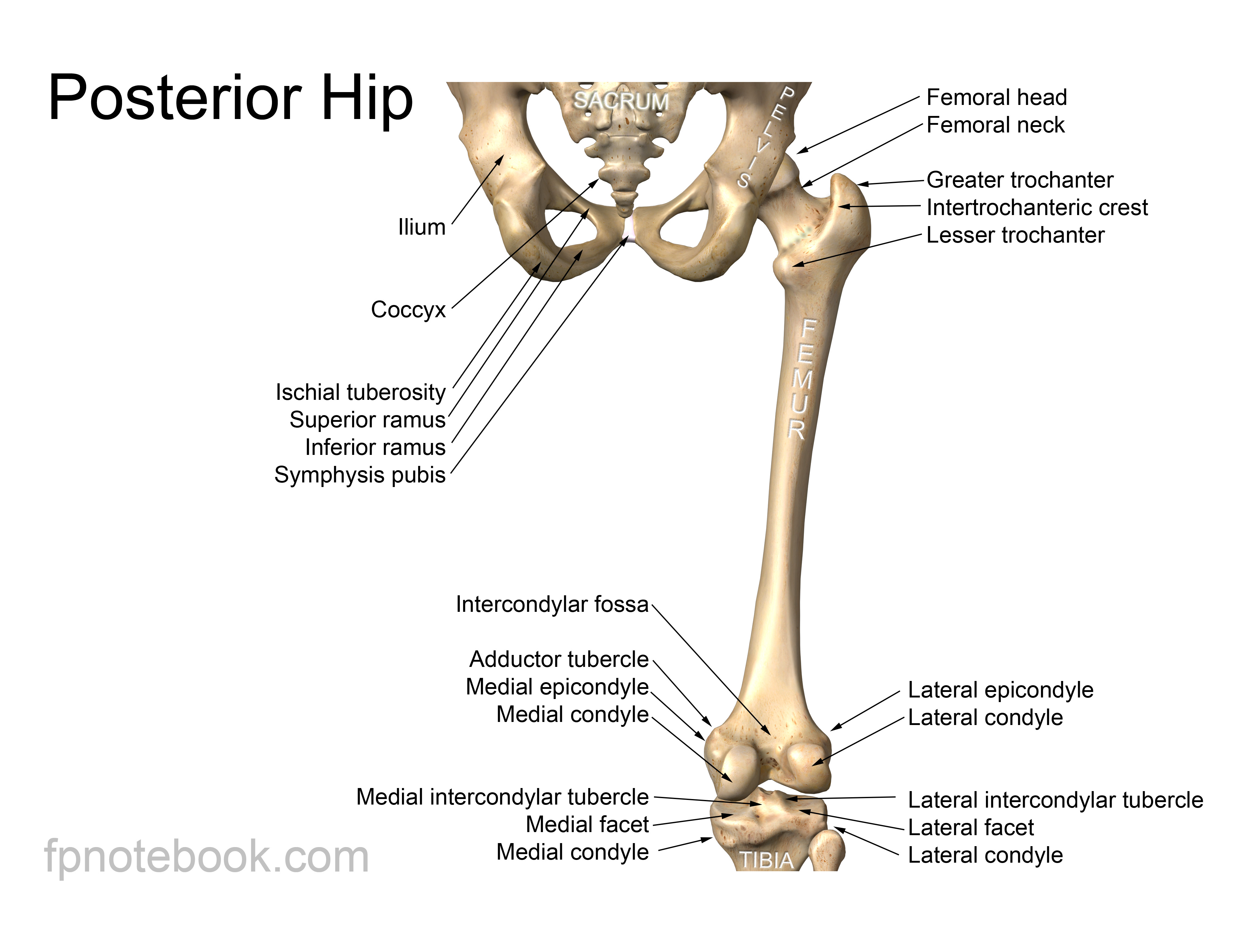
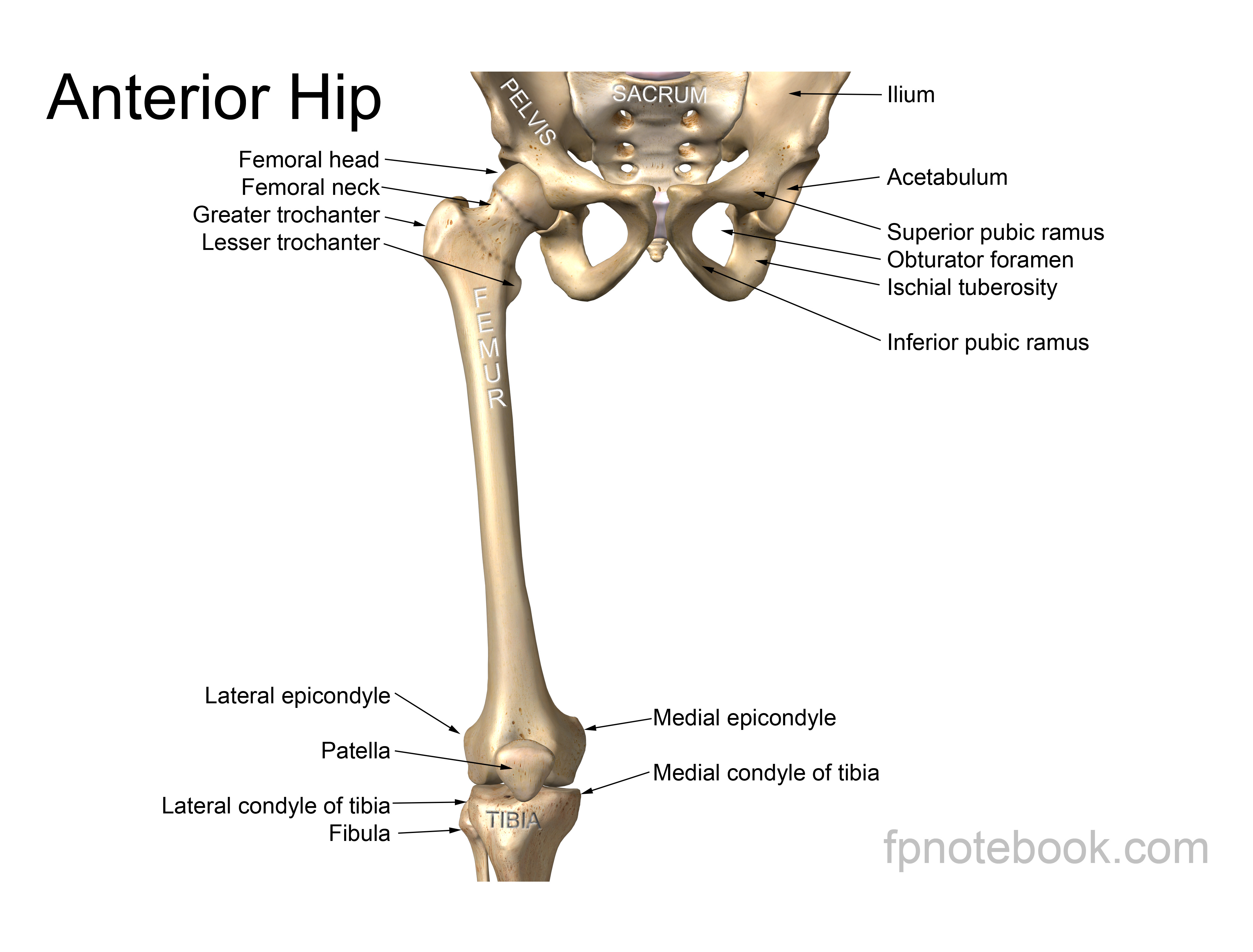
- Bony Landmarks
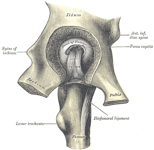
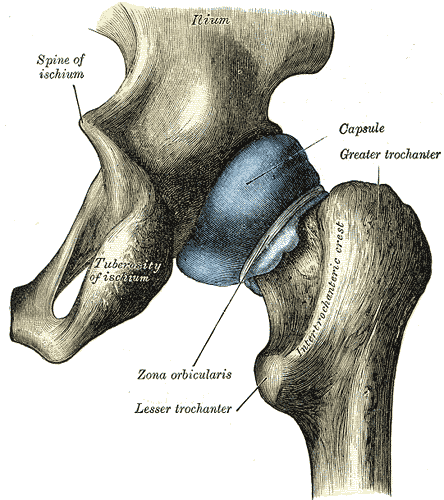
- Hip Joint (ball and socket joint)
- Femoral head articulates with the pelvic acetabulum
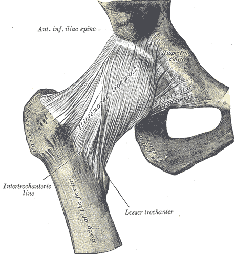
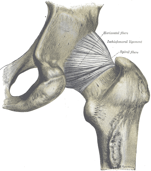
- Hip Joint capsule
- Encapsulates and supports the femoral head and neck, including vascular supply via circumflex arteries
- Subcapital Fractures (e.g. Femoral Neck Fractures) disrupt femoral head vascular supply (AVN risk)
- Greater and lesser trochanters (intertrochanteric), shaft and other distal femur structures lie outside the capsule
- Extracapsular Fractures do not affect femoral head vascular supply
- Encapsulates and supports the femoral head and neck, including vascular supply via circumflex arteries
- Hip Bones (Innominate Bones or Os Coxae)
- Bony Pelvis
- Left and Right Pelvic Bones (ilium, Ischium and Pubis) join at Symphysis Pubis anteriorly
- Sacrum joins each Pelvic Bone posteriorly
- Hip Joint (ball and socket joint)
- Contents
III. Anatomy
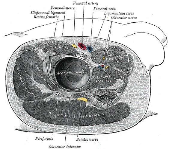
- Hip Images
 Also available as a Poster size image. See printing instructions and image restrictions.
 Also available as a Poster size image. See printing instructions and image restrictions.
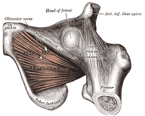
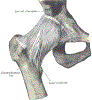 Lewis (1918) Gray's Anatomy 20th ed (in public domain at Yahoo or BartleBy)
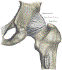 Lewis (1918) Gray's Anatomy 20th ed (in public domain at Yahoo or BartleBy)
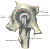 Lewis (1918) Gray's Anatomy 20th ed (in public domain at Yahoo or BartleBy)
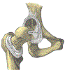 Lewis (1918) Gray's Anatomy 20th ed (in public domain at Yahoo or BartleBy)
 Lewis (1918) Gray's Anatomy 20th ed (in public domain at Yahoo or BartleBy)
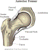
 Lewis (1918) Gray's Anatomy 20th ed (in public domain at Yahoo or BartleBy)
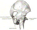 Lewis (1918) Gray's Anatomy 20th ed (in public domain at Yahoo or BartleBy)
-
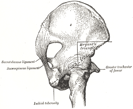
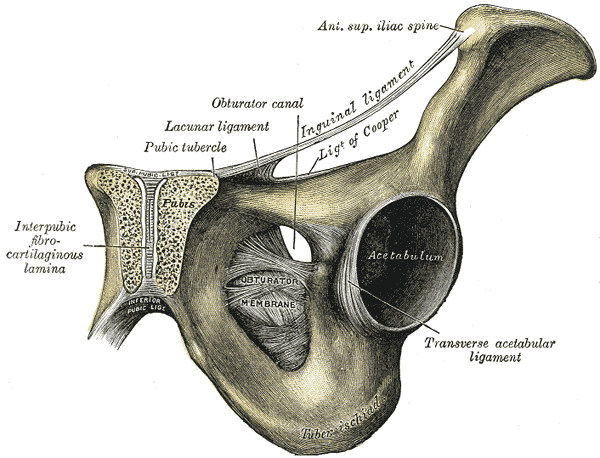
Pelvis Images
 Lewis (1918) Gray's Anatomy 20th ed (in public domain at Yahoo or BartleBy)
 Lewis (1918) Gray's Anatomy 20th ed (in public domain at Yahoo or BartleBy)
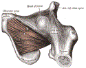 Lewis (1918) Gray's Anatomy 20th ed (in public domain at Yahoo or BartleBy)


