
II. Background
- When referring to anatomic positions within the hand (similar to hand)
- Use radial (instead of lateral) and ulna (instead of medial)
- Use volar (instead of anterior) and dorsal (instead of posterior)
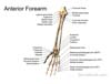
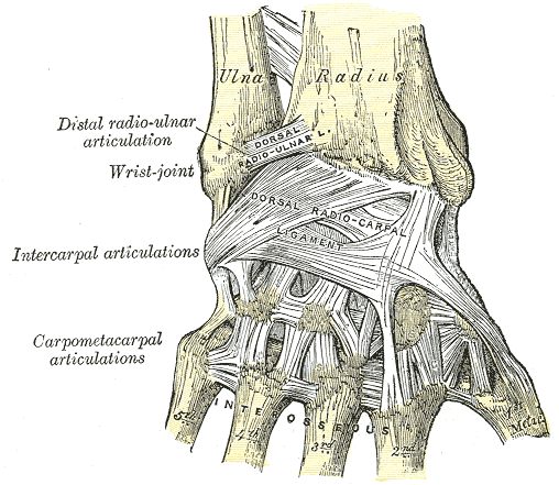
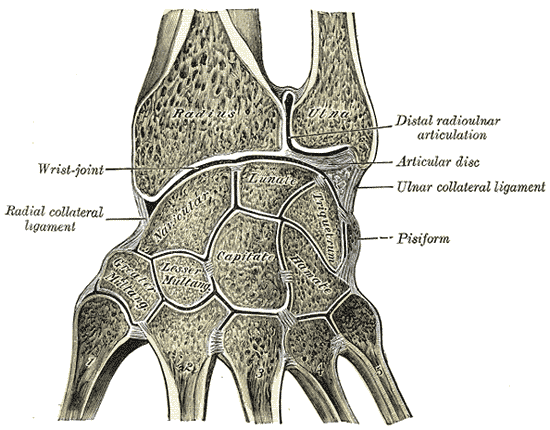
III. Anatomy: Bones and Ligaments
-
 Also available as a Poster size image. See printing instructions and image restrictions.
-
 Also available as a Poster size image. See printing instructions and image restrictions.
-

-
 Lewis (1918) Gray's Anatomy 20th ed (in public domain at Yahoo or BartleBy)
-
 Lewis (1918) Gray's Anatomy 20th ed (in public domain at Yahoo or BartleBy)
-
 Lewis (1918) Gray's Anatomy 20th ed (in public domain at Yahoo or BartleBy)


IV. Anatomy: Muscles
-
 Lewis (1918) Gray's Anatomy 20th ed (in public domain at Yahoo or BartleBy)
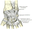
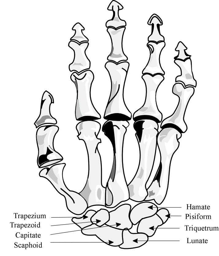
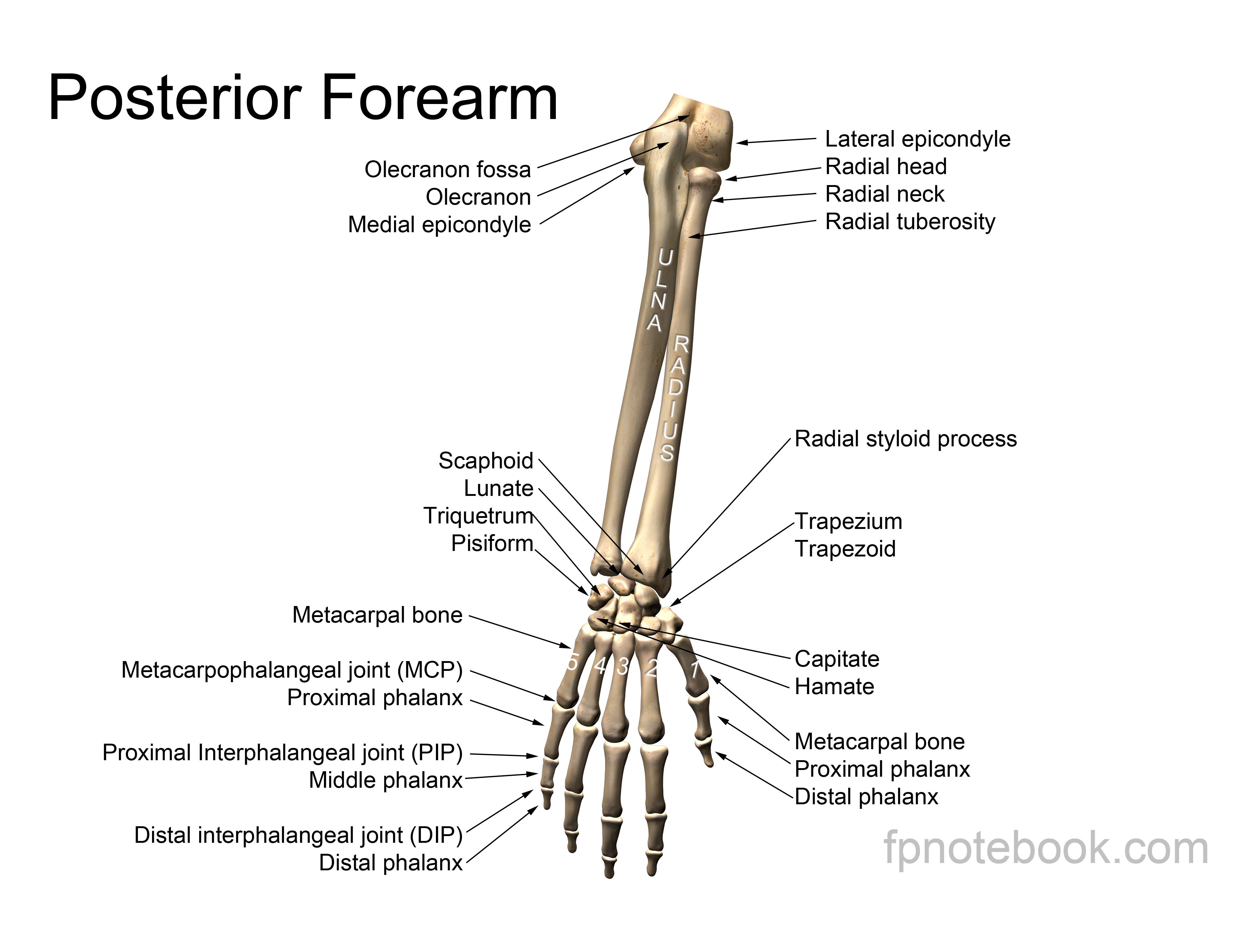
V. Anatomy: Bones
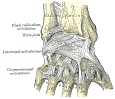
VI. Anatomy: Wrist Bones Mnemonic
-
General
- Two rows of 4 Carpal Bones each (8 total)
- Carpal Bones are strongly linked to one another by ligaments
- Proximal Row (Radial to ulnar wrist)
- Scaphoid or Carpal Navicular (Some)
- Links proximal to distal carpal row
- Lunate (Lovers)
- Triquetral (Try)
- Pisiform (Positions)
- Scaphoid or Carpal Navicular (Some)
- Distal Row (Radial to ulnar wrist)
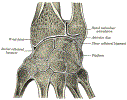
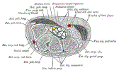
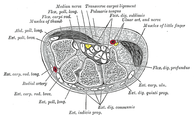
VII. Anatomy: Other Wrist Structures
-
Carpal Tunnel
- See Carpal Tunnel Syndrome
- Finite space bordered by transverse carpal ligament (Flexor Retinaculum)
- Components of Carpal Tunnel
- Finger flexors (9 tendons) course through tunnel
- Median Nerve
- Triangular Fibrocartilage Complex (TFCC)
- Articular disc
- Divides ulna from proximal carpal row
- Injured in Triangular Fibrocartilage Complex Injury
VIII. References
- Tubbs and Janicki (2025) Wrsit XRay, Mastering Emergency Imaging, CCME, accessed 2/15/2026