
II. Images
III. Preparation
- Consider using acoustic stand-off pad on Ultrasound probe (or excessive Ultrasound gel)
- Linear Ultrasound probe
IV. Technique: Dorsal Wrist
- Positioning
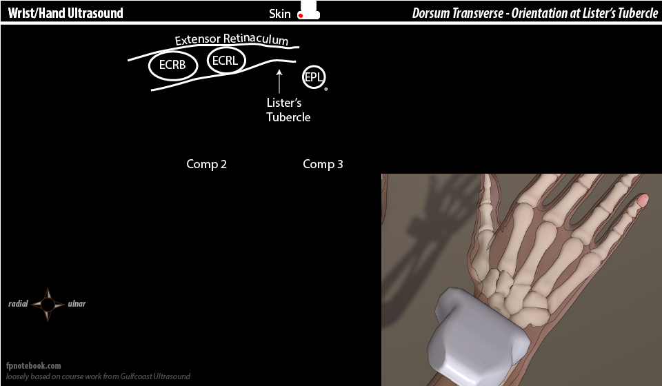
- View 1: Extensor (dorsal) compartments 1-3 in Short Axis (SAX)
- Ultrasound probe
- Probe indicator towards radial aspect of wrist (radius at left screen)
- Slide towards radial aspect to view the first dorsal compartment after identifying Lister's Tubercle
- Images
- Components
- Extensor compartment 1 (lateral aspect of radius near snuff box)
- Abductor Pollicis Longus (APL)
- Extensor Pollicis Brevis (EPB)
- Extensor compartment 2 (dorsal aspect of radius)
- Extensor Carpi Radialis Longus (ECRL)
- Extensor Carpi Radialis Brevis (ECRB)
- Lister's Tubercle
- Extensor Compartment 3
- Extensor Pollicis Longus
- Extensor compartment 1 (lateral aspect of radius near snuff box)
- Ultrasound probe
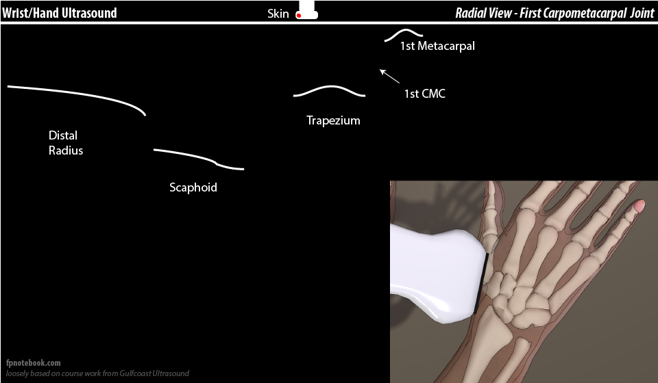
- View 2: First Carpometacarpal Joint at dorsolateral wrist in long axis (LAX)
- Ultrasound probe
- Probe in long axis, with indicator towards proximal arm and elbow
- Start with probe over distal radius and slide distally as Scaphoid, Trapezium and First Metacarpal come into view
- Images
- Components (from screen left to right)
- Distal Radius
- Scaphoid
- Trapezium
- First carpometacarpal joint (CMC-1)
- May be used to direct needle for CMC-1 Injection
- First Metacarpal
- Additional views: De Quervains Tenosynovitis
- Slide probe over the abductor pollicis longus (APL) and extensor pollicis brevis (EPB) in long axis
- Probe may be rotated 90 degrees to view the APL and EPB in short axis
- Ultrasound probe
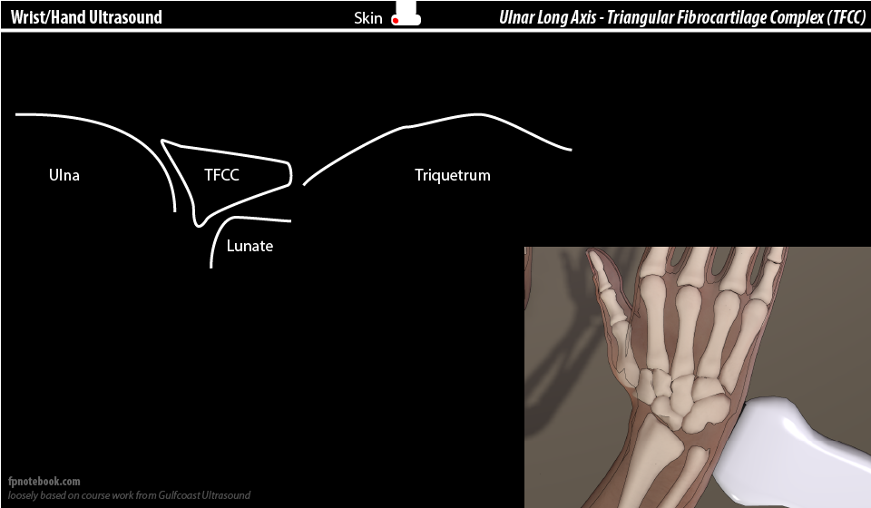
- View 3: Dorsal-medial wrist (ulnar wrist and TFCC)
- Positioning and Ultrasound probe
- Probe in long axis, with indicator towards proximal arm and elbow
- Patient radially deviates the wrist to open the ulnar aspect of the wrist
- Images
- Components
- Distal Ulna
- Triangular Fibrocartilage Complex (TFCC)
- Represents only the TFCC homologue (most superficial aspect, most remains hidden within joint)
- Lunate (deep to TFCC)
- Triquetrum
- Positioning and Ultrasound probe
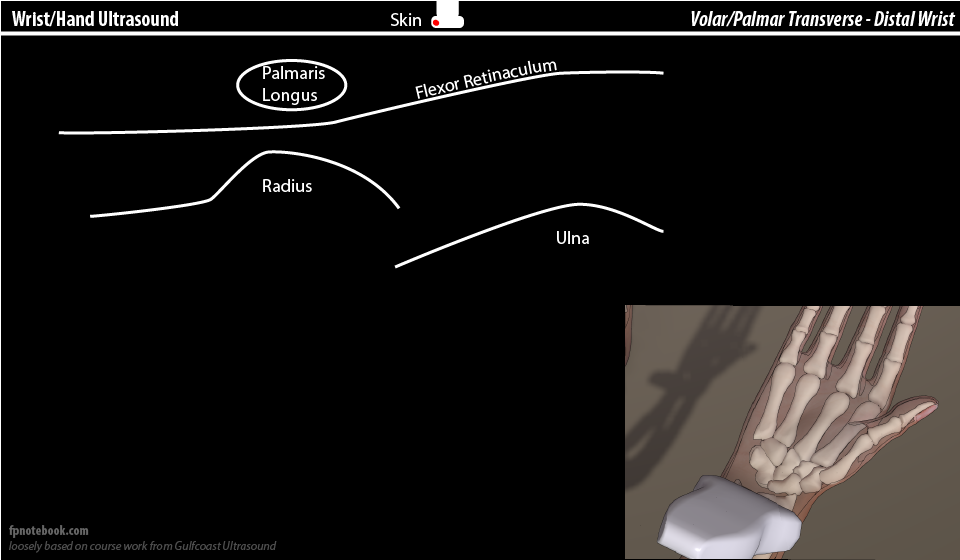
V. Technique: Volar Aspect
VI. References
- Moore (2016) GCUS Musculoskeletal Ultrasound Course, St. Pete's Beach, FL
- Moore (2013) Upper Extremity Ultrasound Video, Gulf Coast Ultrasound