
II. Indication
IV. Anatomy
- See Lumbar Spine Anatomy
- Thoracic Spine Vertebrae are interpreted in similar fashion to lumbar Vertebrae
- Images
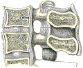
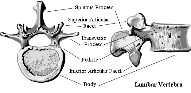
- Lumbar SpineVertebra cross section and lateral view
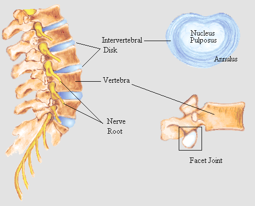
- Lumbar Spine nerve roots and intervertebral discs
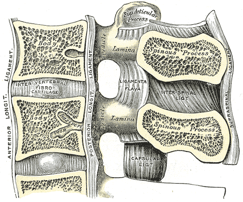
- Spinal Ligaments
 Lewis (1918) Gray's Anatomy 20th ed (in public domain at Yahoo or BartleBy)
- Lumbar SpineVertebra cross section and lateral view
V. Views: Standard (Includes lower thoracic Vertebrae to Sacrum)
- Anterior-Posterior (AP) L-S Spine
- Appears as a totem pole of vertically stacked owl heads
- Vertebral body (owl head)
- Pedicles (owl eyes)
- Spinous process (owl nose)
- Transverse process (owl ear)
- Vertical alignment of spinous processes
- Rotational injury (unilateral facet dislocation)
- Scoliosis
- Other Vertebral injury findings
- Lumbar Spine imaging Includes Pelvis
- Acetabulum and femoral heads
- Pelvis degenerative changes
- Appears as a totem pole of vertically stacked owl heads
- Lateral L-S Spine
- Landmarks
- Alignment and structure
- Lordotic curvature of L-S Spine
- Vertebral alignment
- Findings
- Spondylolisthesis (anterior slippage of L5 on S1)
- Intervertebral disc space height shortening
- Lumbosacral disc disease (degenerative)
- Spinal Osteomyelitis
VI. Views: Specifically Indicated Views
- Flexion and extension views
- Used to assess ligamentous and bony injury
- Contraindications
- Other XRay abnormalities are present
- Decreased Level of Consciousness
- Oblique L-S Spine
- Tube angled at 45 degrees (midway between an AP and Lateral view)
- Interpretation
- Oblique spine view appears as Scotty Dogs stacked on top of one another
- Scotty Dog appearance of the posterior Vertebra overlapping the Vertebral body
- Scotty Dog oriented with the buttocks to the right side
- Demonstrates the right lamina, pedicle and pars interarticularis
- Scotty Dog oriented with the buttocks to the left side
- Demonstrates the left lamina, pedicle and pars interarticularis
- Scotty Dog
- Inferior intervertebral articular processes (Scotty Dog's front and hind legs)
- Superior intervertebral articular processes (Scotty Dog's ears and tail)
- One lamina (Scotty Dog's body)
- One pedicle (Scotty Dog's eye)
- One transverse process (Scotty Dog's nose)
- One Pars interarticularis (Scotty Dog's neck)
- Scotty Dog with a collar (Fracture line), suggests a Pars Defect (Spondylolysis)
- Pars Defect on both sides is associated with Spondylolisthesis
- Oblique spine view appears as Scotty Dogs stacked on top of one another
- Findings/Indications (has been replaced by CT and MRI)
- Neural foramina narrowing
- Pars Interarticularis Defect
- Spinal Tumor
- Facet hypertrophy
- Spondylosis
- Spondylolysis
- Spondylolisthesis
- Resources
- Oblique Lumbar Spine View (Radiopaedia)