
II. Views
- Standard Views
- Anteroposterior
- Epicondyles (medial and lateral)
- Humeral capitellum and trochlea
- Radial head
- Ulna's coronoid process
- Lateral
- True lateral view
- Lateral and medial condyles should overlap
- Figure of 8 (hour glass, or snowman)
- Bottom of the figure 8 represents overlapping distal humeral capitellum and trochlea
- Landmarks include radial head and olecranon
- Posterior olecranon fossa (posterior fat pad)
- Anterior coronoid fossa (anterior fat pad and Sail Sign)
- True lateral view
- Anteroposterior
- Special views
- Radial Head-Capitellum View (oblique view)
- Isolates radial head without overlapping shadows
- Radial Head-Capitellum View (oblique view)
III. Technique
- Elbow in 90 degree flexion
- Compare with opposite elbow
IV. Evaluation: Landmarks on lateral Elbow XRay
- Anterior humeral line
- Line drawn along the anterior edge of the Humerus
- Passes through middle third of the capitellum
- Radiocapitellar Line
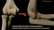
- Line drawn longitudinally through center of proximal radius
- Line should bisect the capitellum (or through the middle third of the capitellum)
V. Evaluation: Findings suggestive of Fracture
- Background
- Anterior and posterior fat pads fill the normal elbow recesses
- Anterior: Coronoid fossa
- Posterior: Olecranon fossa
- When injury or inflammation results in a hemarthrosis or joint effusion, the fat pads are displaced outwards
- XRay with dark fat overlying lighter appearing soft tissue results in fat pad related signs
- Anterior fossa is more shallow, displacing its fat pad with less fluid than the posterior fossa
- Posterior fat pad is always abnormal
- Anterior and posterior fat pads fill the normal elbow recesses
- Sail Sign
- Effusion displaces the anterior fat pad forming a dark triangle in the anterior fossa
- Consider Radial Head Fracture
- May also represent synovitis or spontaneous hemarthrosis without Fracture
- Small anterior fat pads that hug the bone (no "sail") are typically normal and physiologic
- Posterior fat pad sign (always pathologic)
- Child: Supracondylar Fracture
- Adult: Radial Head Fracture
VI. Findings
VII. Precautions
-
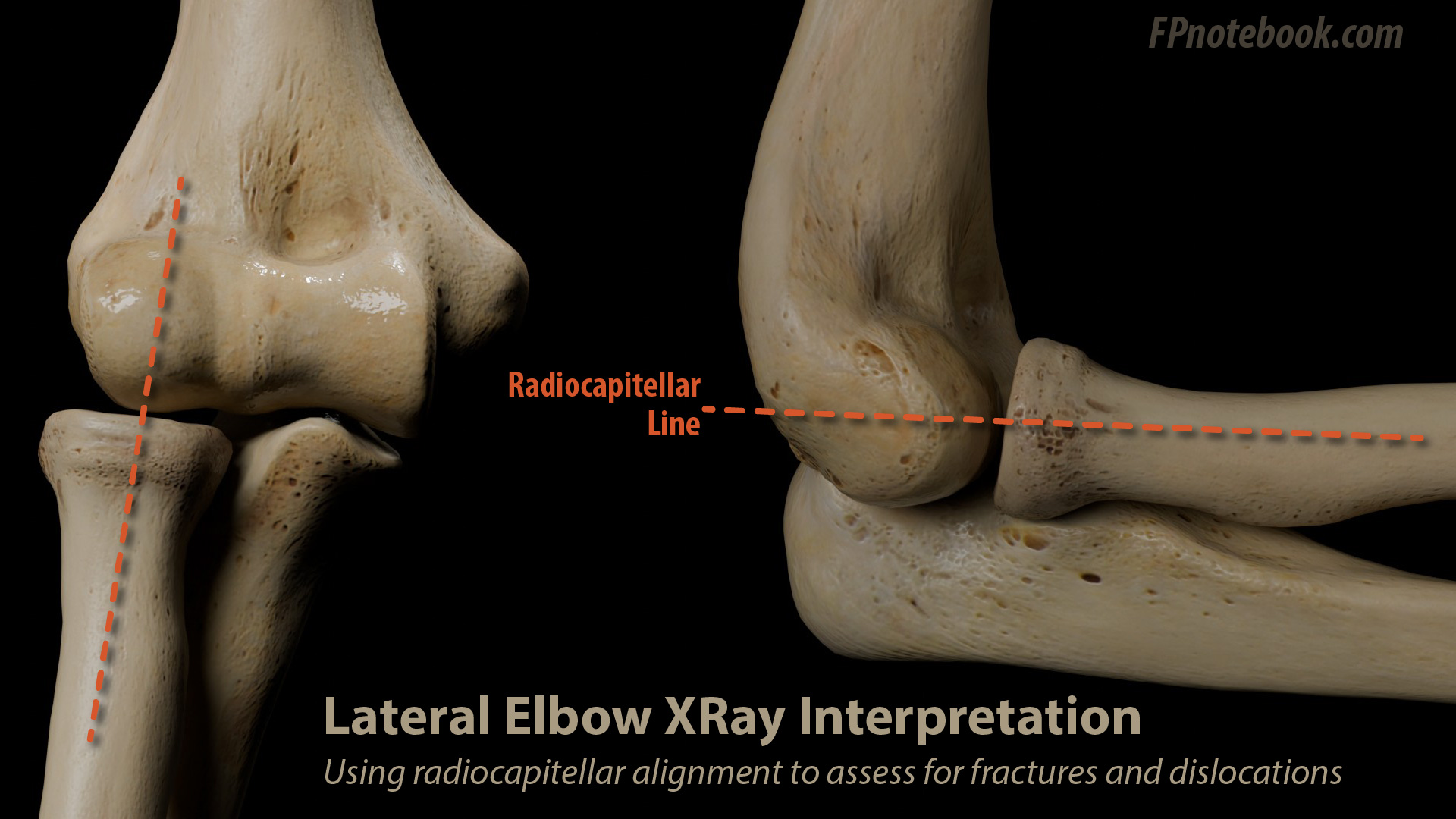
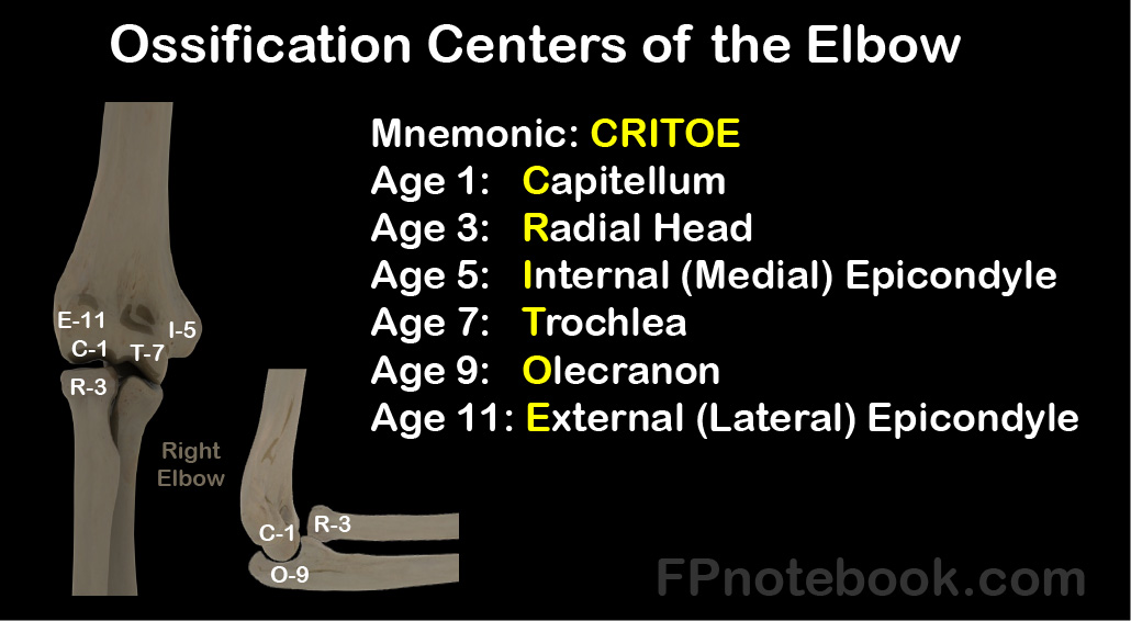
Elbow Ossification Centers
-
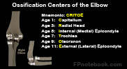
- Consider when reviewing Elbow XRays in children
-
VIII. References
- Gharahbaghian in Herbert (2017) EM:Rap 17(11): 7-9