
II. Physiology
- Elbow is a hinged joint with the ability to pronate and supinate
- Elbow is comprised of 3 joints articulating between the distal Humerus condyles and the radius and ulna
- Ulno-humeral joint
- Distal Humerus (trochlea) articulates with the proximal ulna (trochlear notch)
- Trochlea notch lies between the ulna's olecranon proximally/posteriorly and the coronoid process distally
- Radio-capitellar joint
- Distal Humerus (capitellum) articulates with the radial head
- Radio-ulnar joint
- Radial head articulates with the ulna
- Allows for Forearm supination and pronation
- Ulno-humeral joint
- Collateral ligaments
- Medial and Lateral collateral ligaments add elbow joint stability
- Olecranon bursa
- Allows skin to course over the surface of the olecranon smoothly during elbow rotation
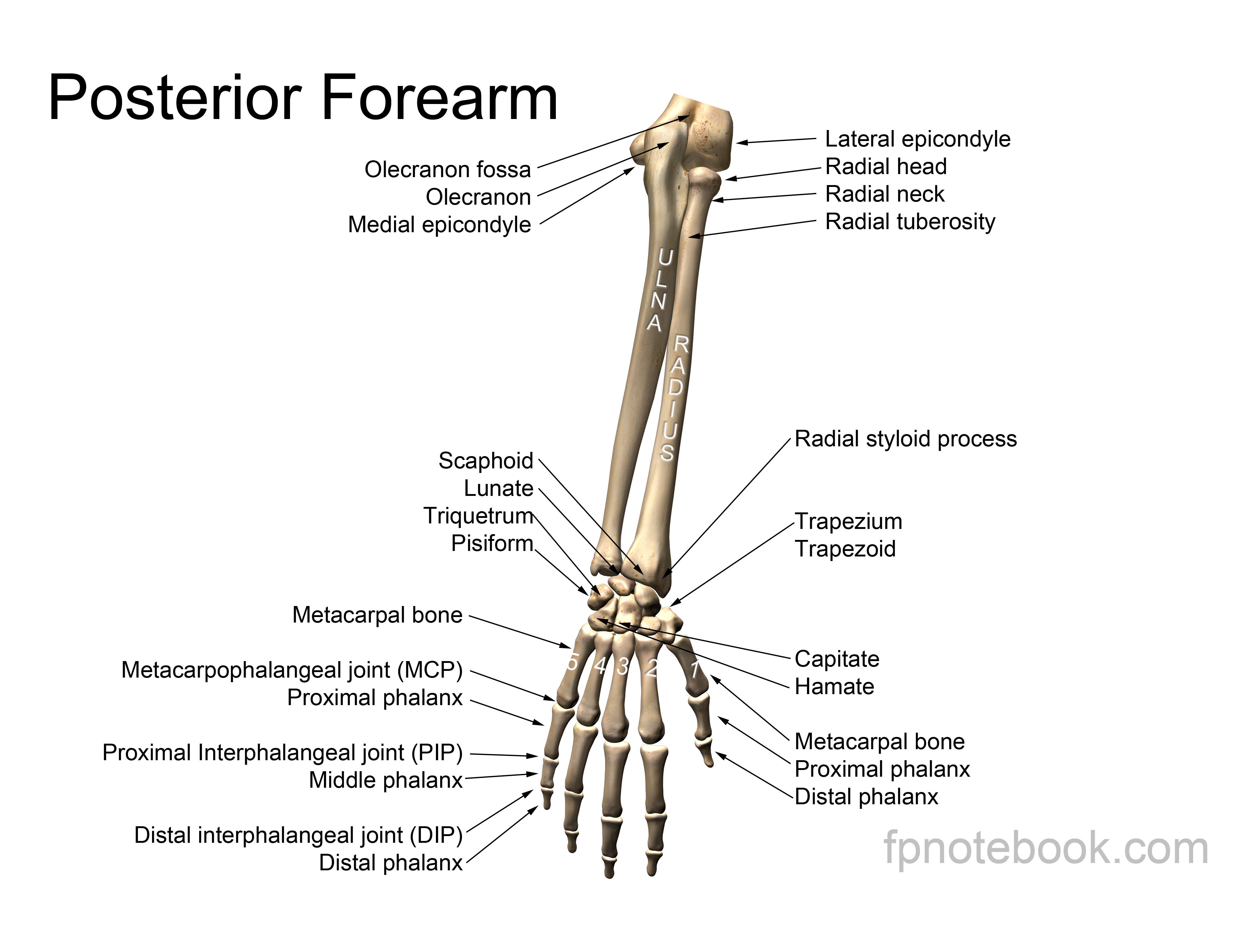
III. Anatomy: Bones and Ligaments
-
General
- Bony Landmarks form a triangle (with elbow at 90 degrees)
- Olecranon
- Lateral epicondyle
- Medial epicondyle
- Bony Landmarks form a triangle (with elbow at 90 degrees)
-
Humerus
- Widens distally forming lateral and medial epicondyles
- Radial Head
- Articulates with capitellum (at lateral epicondyle)
- Articulates with lateral ulna
- Held in position by orbicular ligament
- Easily palpable near lateral epicondyle
- Ulna
- Articulates with Trochlea (at Medial epicondyle)
- Epicondyles
- Images
 Also available as a Poster size image. See printing instructions and image restrictions.
 Also available as a Poster size image. See printing instructions and image restrictions.
 Lewis (1918) Gray's Anatomy 20th ed (in public domain at Yahoo or BartleBy)
 Lewis (1918) Gray's Anatomy 20th ed (in public domain at Yahoo or BartleBy)
 Lewis (1918) Gray's Anatomy 20th ed (in public domain at Yahoo or BartleBy)
 Lewis (1918) Gray's Anatomy 20th ed (in public domain at Yahoo or BartleBy)
 Lewis (1918) Gray's Anatomy 20th ed (in public domain at Yahoo or BartleBy)
 Lewis (1918) Gray's Anatomy 20th ed (in public domain at Yahoo or BartleBy)


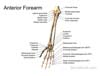
IV. Anatomy: Muscles and Tendons
- Tendon insertions
- Triceps attaches to Olecranon posteriorly
- Biceps and brachialis attach to radius and ulna
- Pronators and Supinators Mnemonic
- MFP: medial (epicondyle) flexors and pronators
- LES: lateral (epicondyle) extensors and supinators
- Images
 Lewis (1918) Gray's Anatomy 20th ed (in public domain at Yahoo or BartleBy)

V. Anatomy: Nerves
-
General
- Median and Radial Nerves (as well as the brachial artery) are contained within the Antecubital Fossa
- Ulnar Nerve in contrast, courses posterior to the medial epicondyle
-
Median Nerve
- Deep in antecubital fossa
- Medial to biceps
-
Radial Nerve
- Lateral to biceps and brachialis Muscles
-
Ulnar Nerve
- Posterior to medial epicondyle
- In groove between medial epicondyle and Olecranon
- Superficial, and vulnerable to injury
VI. Anatomy: Vascular
- See Vascular Anatomy of the Elbow
- Artery
- Vein
 Lewis (1918) Gray's Anatomy 20th ed (in public domain at Yahoo or BartleBy)