
II. Images

III. Technique: Anterior Elbow
- Positioning
- Patient lies supine with arm resting at side
- Elbow slightly flexed and wrist supinated (consider towel roll under wrist)
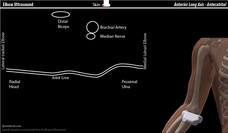
- View 1: Antecubital space (probe transverse or short axis, SAX)
- Landmarks
- Humerus capitulum and trochlea form dual parallel lines with posterior shadowing
- Mnemonic: Pyramid
- Bicipital tendon sits atop the pyramid centrally
- "Pyramid walls" contain the brachialis Muscle
- Draw a lateral pyramid wall between bicipital tendon to the radial head (or humeral capitulum)
- Draw a medial pyramid wall between bicipital tendon to the ulnar notch
- Brachialis Muscle pyramid is flanked by the brachioradialis Muscle laterally (radial aspect)
- Radial Nerve sits between the Brachialis Muscle and the brachioradialis Muscle
- Brachialis Muscle pyramid is flanked by the pronator teres medially (ulnar aspect)
- Brachial artery and Median Nerve are located in this region
- Images
- Components
- Lateral (radial aspect of volar arm)
- Brachioradialis Muscle
- Radial Nerve (embedded in fascia)
- Mid
- Medial (ulnar aspect of volar arm)
- Brachial artery (most superficial)
- Pronator teres
- Median Nerve (embedded in fascia, just medial and deep to the brachial artery)
- Trochlea of Humerus (articulates with ulnar coronoid anteriorly and olecranon posteriorly)
- Lateral (radial aspect of volar arm)
- Structures that may be followed distally (rotate to long access)
- Bicipital tendon (to its distal two part insertion)
- Radial Nerve (into two branches)
- Median Nerve
- Landmarks
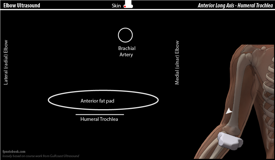
- View 2: Antecubital Space short axis (SAX) tilted slightly cephalad from view 1
- Positioning
- Patient flexes elbow to 90 degrees
- Images
- Landmarks
- Brachial artery (superficial)
- Anterior fat pad
- Humeral coronoid fossa
- Significance
- Anterior fat pad displacement (Fracture) is more prominent on Ultrasound than Sail Sign on xray
- Positioning
IV. Technique: Bicipital tendon visualization
- View 1: Anterior long axis
- Follow bicipital tendon from distal Humerus region and antecubital space to dual proximal radius insertion sites
- View 2: Medial long axis or Pronator Window (most reliable)
- Position elbow flexed to 90 degrees
- Start distal to medial epicondyle in long axis
- Gradually slide the probe anteriorly until brachial artery is visible in long axis
- Bicipital tendon will run in parallel, immediately deep to the brachial artery
- View 3: Lateral long axis
- Position elbow flexed to 90 degrees
- View 4: Posterior long axis (dorsal approach)
- Best for distal bicipital tendon visualization (last 1-2 cm) and injection
- Position elbow flexed to 90 degrees
- Pronate the Forearm to expose the bicipital tendon
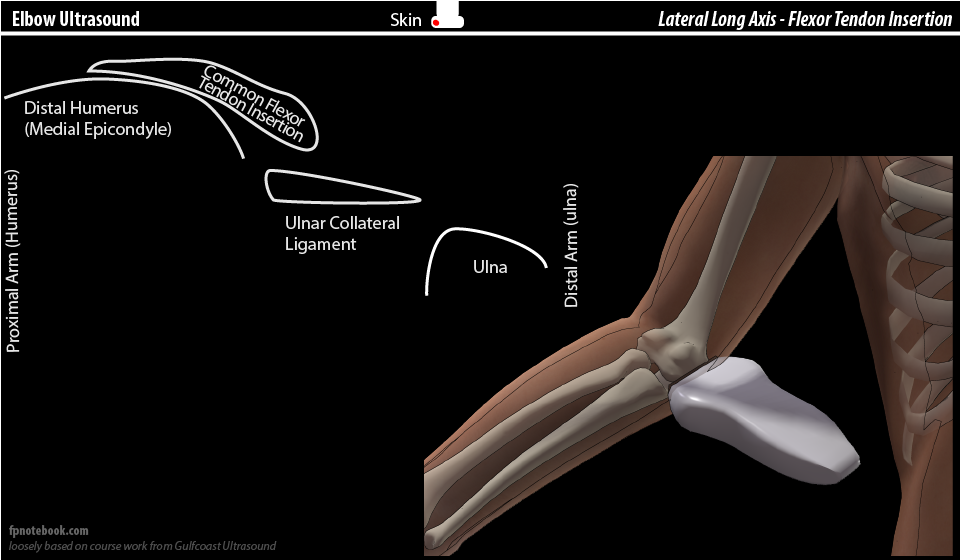
V. Technique: Medial View
- Patient Positioning
- Elbow extended and Forearm supinated (thumb lateral)
- Ultrasound probe indicator toward Shoulder (left image is proximal, toward Shoulder)
- Medial Elbow Image (Medial Epicondyle)
- View: Long Axis (LAX) of medial elbow
- Superficial structures
- Deep structures
- Distal Humerus (medial epicondyle proximal to trochlea)
- Ulnar collateral ligament (triangular)
- May require increased downward probe pressure and probe rotation for better visualization
- Joint line (gap between trochlea and ulna)
- Proximal ulna (deeper)
VI. Technique: Lateral View
- Patient Positioning
- Patient sitting or lying, elbow flexed 60-90 degrees and Forearm pronated (palm down, thumb medial)
- Ultrasound probe indicator toward Shoulder (left image is proximal, toward Shoulder)
- Scan plane should be lateral to medial (parallel to floor, aiming towards medial epicondyle)
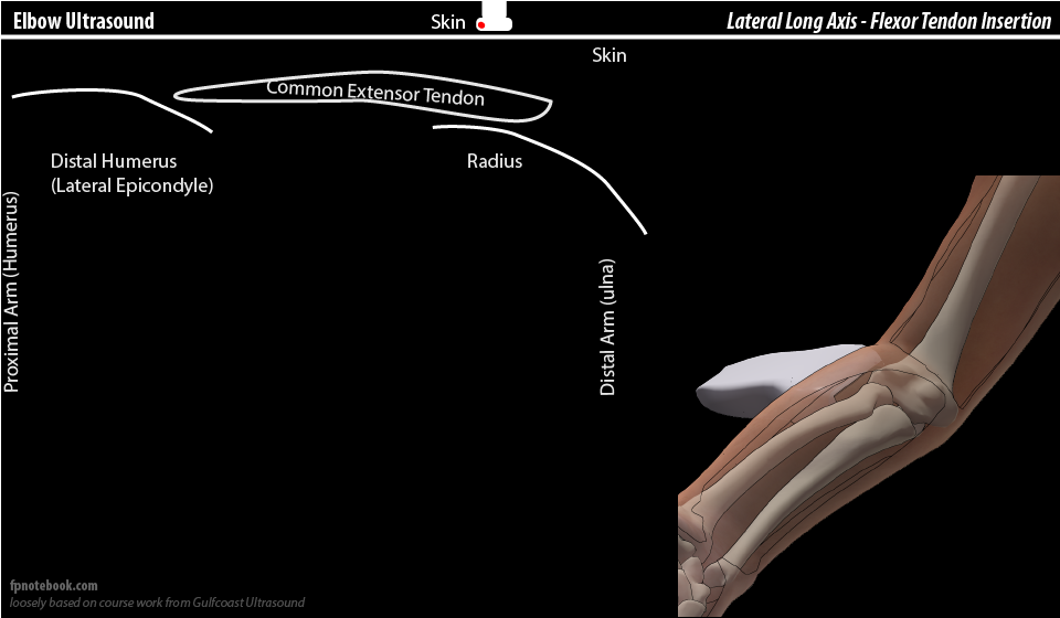
- Lateral Elbow Image (Lateral Epicondyle)
- View: Long Axis (LAX) of lateral elbow
- Humerus (lateral epicondyle, capitellum)
- Common extensor tendon (superficial, inserts on lateral epicondyle)
- Joint space
- Radius (radial head)
- Rotation visible on supination and pronation dynamic maneuvers
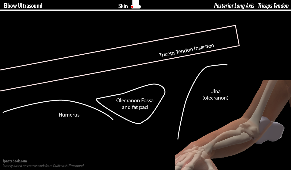
VII. Technique: Posterior View
- Patient Positioning
- Patient sitting with hand pronated, palm on hip (thumb medial) and elbow flexed posteriorly (crab position) OR
- Patient lying with elbow flexed and propped on pillow
- View 1: Short Axis (SAX) of Ulnar Nerve in ulnar groove (posterior-medial elbow)
- Humerus (Medial Epicondyle)
- Ulnar groove
- Ulnar Nerve (normal cross sectional area: 7mm)
- Nerve may be tracked in short axis
- Ulna (Olecranon)
- View 2: Long Axis (LAX) of Triceps Muscle, tendon and insertion at ulna (medial olecranon)
- View 3: Short Axis (SAX) of Posterior Fat Pad (and triceps tendon)
- Triceps tendon (superficial)
- Triceps Muscle (hypoechoic, deeper)
- Olecranon fossa with posterior fat pad
VIII. References
- Jacobson (2013) Musculoskeletal Ultrasound, Elsevier, Philadelphia, p. 72-109
- Moore (2013) Upper Extremity Ultrasound Video, GCUS
- Moore (2015) Sonography of the Extremities, 4th ed, p. 25-34
- Lento (2016) Elbow, GCUS Musculoskeletal Ultrasound Course, St Pete's Beach, attended 1/25/2016