
II. Images
III. Preparation: Ultrasound
- Low frequency (5 MHz) curved array probe
- Depth: Start at 9-10 cm
IV. Technique: Anterior Hip in Long Axis (LAX) and Hip Arthrocentesis
- Indications
- Differentiates hip effusion (intracapsular) from Iliopsoas Bursitis (extracapsular)
- Best image to guide Hip Injection or aspiration (broad femoral neck as target)
- Positioning
- Patient supine with hip in neutral position (hip exposed to inguinal ligament)
- Internal rotation of hip and knee flexion may aid visualization of small hip effusions
- Slight external rotation may help to visualize other structures
- Ultrasound linear probe in line with femoral neck (perpendicular to mid-inguinal ligament)
- Palpate femoral artery and probe is placed lateral to this position
- Ultrasound probe indicator toward Umbilicus
- Patient supine with hip in neutral position (hip exposed to inguinal ligament)
- Images
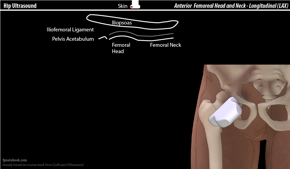
- Components
- Iliopsoas Muscle (overlying hip)
- Overlies all deeper structures
- Acetabulum of hip (screen left)
- Bright, hyperechoic
- Femur with overlying capsule
- Extra-capsular space (Iliopsoas bursa)
- Joint capsule
- Linear band follows femoral head and concave over femoral neck
- Hyperechoic due to contained iliofemoral and pubofemoral ligaments
- Intracapsular space
- Increasing depth (and convex expansion of the space) suggests Hip Joint effusion
- Normal depth: 5 mm or within 1-2 mm of intracapsular space depth of opposite hip
- Abnormal depth >8-9 mm
- Bone
- Femoral head
- Rounded, convex
- Femoral neck
- Femoral head
- Iliopsoas Muscle (overlying hip)
-
Arthrocentesis
- Prepare skin
- Mark landmarks (but also performed under Ultrasound guidance)
- Antiseptic (e.g. Chlorhexidine)
- Local Skin Anesthesia (and along intended track) with Lidocaine 1% with Epinephrine
- Aspirate joint
- Spinal needle (3.5 inch, 20 gauge) advanced under Ultrasound guidance toward fluid pocket
- Prepare skin
V. Technique: Anterior Hip in Transverse or Short Axis (SAX)
- Indications
- Confirms hip effusion (intracapsular)
- Positioning
- Patient supine with hip in neutral position or slight external rotation (hip exposed to inguinal ligament)
- Probe in short axis with approximately 20 degrees angulation (probe indicator toward iliac crest)
- Probe rotated 90 degrees from the long axis view used above
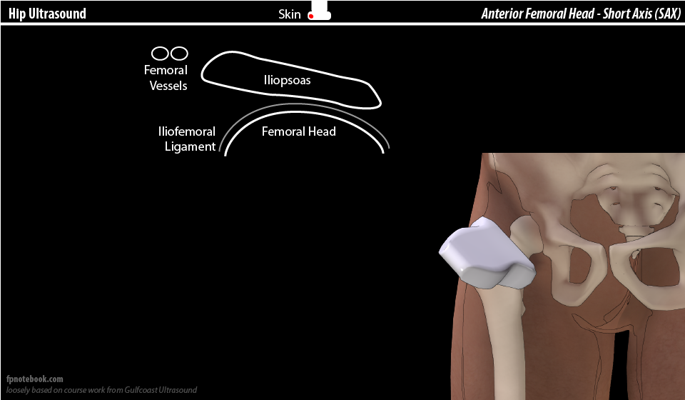
- Images
- Components: Superficial
- Components: Deep to Iliopsoas Muscle
- Joint capsule
- Intracapsular space
- Femoral head
VI. Technique: Lateral Hip at Trochanteric Bursa
- Indications
- Evaluates Trochanteric Bursitis (Gluteus Medius bursa)
- Directs trochanteric bursa region injection (typically tendonosis rather than the actually rare Bursitis)
- Positioning
- Patient lies on their side (decubitus)
- View 1: Long Axis (LAX)
- Positioning
- Ultrasound probe in long axis overlying greater trochanter
- Ultrasound beam tilted slightly anteriorly
- Better demonstrates IT Band interface
- Gluteus minimus attachment at anterior facet
- Ultrasound beam tilted slightly anteriorly
- Gluteus medius attachment at lateral facet
- Components
- Gluteus Maximus and Iliotibial Band (superficial)
- Joint capsule
- Femoral Greater Trochanter
- Positioning
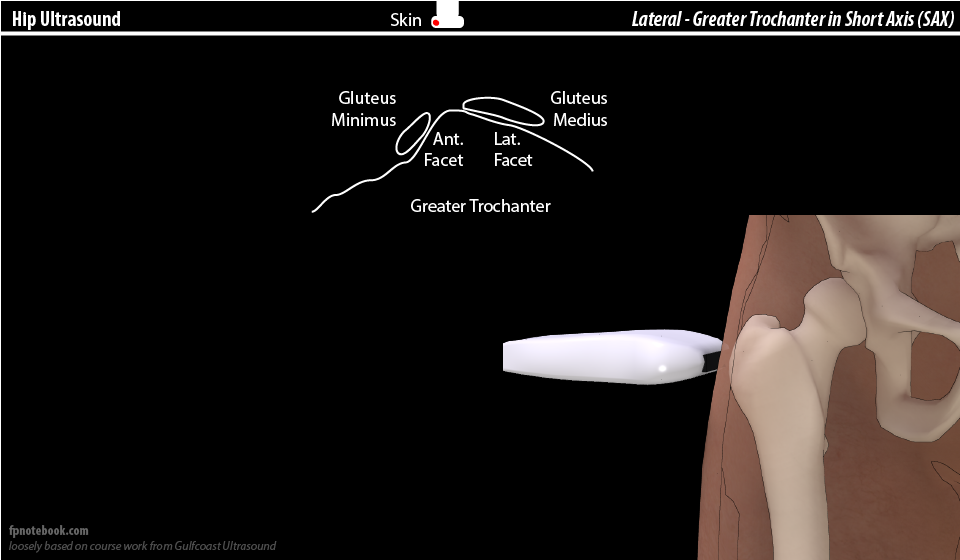
- View 2: Short Axis (SAX, transverse)
- Positioning
- Ultrasound probe in short axis overlying greater trochanter (probe turned 90 degrees from above)
- Images
- Components: Deep
- Anterior facet of Greater trochanter
- Appears similar to the front facing side of half dome (Yosemite)
- Gluteus minimus attaches at anterior facet
- Lateral facet of Greater trochanter
- Appears similar to the back side of half dome (Yosemite)
- Gluteus medius attaches at lateral facet
- Anterior facet of Greater trochanter
- Positioning
VII. Technique: Posterior Hip at Piriformis Muscle
- Indications
- Piriformis injection in Sciatica (or Piriformis Syndrome)
- Positioning
- Patient supine
- Ultrasound probe
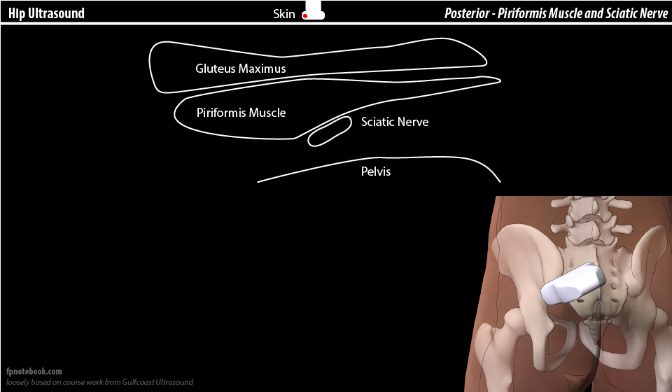
- Images
- Components
- Gluteus maximus Muscle
- Superficial, hypoechoic, large
- Piriformis Muscle
- Deeper, more hyperechoic, small
- Tapers laterally and inserts on greater trochanter
- Identify piriformis Muscle dynamically by rotating foot internally and externally while Ultrasounding
- Sciatic nerve
- Immediately deep to piriformis Muscle
- Gluteus maximus Muscle
VIII. References
- Moore (2010) Hip and Spine Ultrasound Video, GCUS
- Moore (2016) Musculoskeletal Ultrasound Course, Gulf Coast Ultrasound, St. Pete's Beach, FL