
II. Background: Spinal Cord
- Spinal Cord contains two main sets of pathways: Ascending (Sensory) and Descending (Motor).
- Both sensory and Motor Nerves Synapse with other nerves multiple times as they traverse the Central Nervous System.
- Ascending (Sensory)
- Carry sensory signals (pain and Temperature, position sense or proprioception, and light touch)
- Via Peripheral Nerves, past a sensory Ganglion and into the posterior horn of the central cord grey matter
- Follow one of three main ascending pathways, specific for the type of sensory input.
- Descending (Motor).
- Signals via the Corticospinal tract (body) and corticobulbar tract (face)
- Initiated in the Primary Motor Area of the Parietal Lobe and descends the spinal cord
- Exits cord via the anterior root and terminates at the Neuromuscular Junction where it results in muscular contraction
III. Components: Spinal Cord Levels
-
General
- Total spinal nerves: 31 pairs
- Spinal cord ends at L2
- Spinal nerves L2 to S5 descend as individual "horse hairs" (cauda equina)
- Nerves exit at their respective Vertebral levels
-
Cervical Spine
- Cervical spinal nerves: 8 (C1-C8)
- Cervical nerves exit ABOVE their associated Vertebrae
- C1 cervical nerve exits above cervical Vertebra 1 (atlas)
- C7 cervical nerve exits above cervical Vertebra 7
- C8 cervical nerve exits above thoracic Vertebra 1
- Exception, as it is not associated with a cervical Vertebra (only 7 Cervical Vertebrae)
- Marks the transition point, after which all spinal nerves exit below their associated Vertebra
-
Thoracic Spine
- Thoracic spinal nerves: 12 (T1-T12)
- Thoracic nerves exit BELOW the associated Vertebrae
-
Lumbar Spine
- Lumbar spinal nerves: 5 (L1-L5)
- Lumbar nerves exit BELOW the associated Vertebrae
-
Sacrum
- Sacral spinal nerves: 6 (S1-S5 and coccygeal nerve)
IV. Components: Grey matter (central, cell bodies)
- Posterior horn cells (somatic sensory)
- Sensory fibers from the upper tracts Synapse with Lower Motor Neurons (Peripheral Nerves)
- Autonomic or visceral motor nuclei (sympathetic and parasympathetic)
- Visceral Motor Neurons are contained in the middle grey matter (between anterior and posterior horn cells)
- Fibers then course to their visceral targets (e.g. heart, abdominal viscera)
- Anterior horn cells (somatic motor)
- Motor fibers from the Corticospinal tract Synapse with Lower Motor Neurons (Peripheral Nerves)
V. Components: White Matter - Sensory or Ascending Tracts
-
General
- Tracts are named from origin (prefix) to target (suffix)
- All sensory tracts cross the midline, Synapse in the Thalamus and terminate in the contralateral sensory cerebral region
- Exception: Spinocerebellar Tract does not cross the midline
- Spinothalamic Tract (pain and Temperature sense) crosses within a few levels of its spinal cord entry
- Dorsal Columns (proprioception) cross over within the Medulla
- Each Sensation type follows a specific ascending neurologic pathway.
- Pain (nociception) and Temperature sense is carried by the lateral Spinothalamic Tract
- Crude Touch Sensation is carried by the anterior Spinothalamic Tract.
- Conscious Proprioception (position sense or stereognosis, and vibration sense) follows the posterior or Dorsal Columns
- Unconscious proprioception (complex coordination of innate movement such as walking) follows Spinocerebellar Tract
- Light Touch Sensation follows both the Spinothalamic Tract as well as the Dorsal Columns
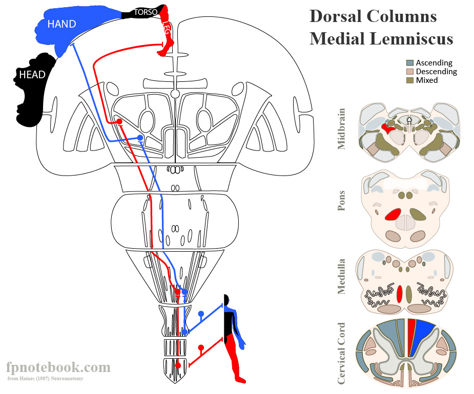
- Posterior Columns or Dorsal Columns (sensory, posterior cord)
- Transmits CONSCIOUS proprioception, vibratory Sensation and stereognosis
- Proprioception senses limb movement and position
- Stereognosis senses an object's identity by touch
- Also partially transmits light Touch Sensation
- Pathway
- Sensory signals rise within the spinal cord from Fasciculus Gracilis and Fasciculus Cuneatus
- Synapse at the Nucleus Gracilis and Nucleus Cuneatus in Medulla to form Internal Arcuate Tract
- Internal Arcuate Tract crosses over the midline at the Medulla to form Medial Lemniscus
- Medial Lemniscus courses to contralateral Thalamus and cerebral cortex
- Fasciculus Gracilis
- Fasciculus Cuneatus
- Spinocervicothalamic Tract
- Located lateral to the Posterior Columns (and to the posterior root entry)
- Transmits sensory signals similar to the typically described Posterior Columns (gracilis and cuneatus)
- Transmits proprioception, stereognosis, vibration (and light touch)
- Images
- Transmits CONSCIOUS proprioception, vibratory Sensation and stereognosis
- Spinocerebellar Tract (sensory, lateral cord)
- Transmits UNCONSCIOUS proprioception Sensation (e.g. walking)
- Does not cross the midline (unlike all other sensory tracts)
- Courses to the ipsilateral (same side) Cerebellum via the superior and inferior peduncles
- Lesion findings
- Ipsilateral Ataxia
- Spinothalamic Tract (sensory, anterior cord)
- Transmits pain and TemperatureSensation
- Also partially transmits light Touch Sensation
- Fibers cross the midline within 1-2 spinal levels of their peripheral Sensory Nerve origin
- Courses to contralateral Thalamus and cerebral cortex (or terminates in Brain Stem)
- Lower extremity fibers are lateral to upper extremity fibers
- With external compression or injury, leg Sensation may be affected earlier than arms
- Images
- Transmits pain and TemperatureSensation

VI. Components: White Matter - Motor or Descending Tracts
- Background
- Tracts are named from origin (prefix) to target (suffix)
- Motor cortex efferent signals are carried via corticospinal (body) and corticobulbar (head) tracts
- Course
- Pass via Internal Capsule and then cross (decussate) in the Medulla
- Descend the spinal cord to specific levels, synapsing at the anterior horn
- Exit the spinal cord, forming Peripheral Nerves and terminating at the Neuromuscular Junctions of Muscles
- Upper Motor Neuron Defects
- Nerve injury occurs above the Peripheral Nerve (e.g. anterior horn, Corticospinal tract)
- Presents with spastic paralysis and hyperreflexia
- Lower Motor Neuron Defects
- Peripheral Nerve Injury
- Presents with Flaccid Paralysis, muscular atrophy, muscle Fasciculations and fibrillations and hyporeflexia
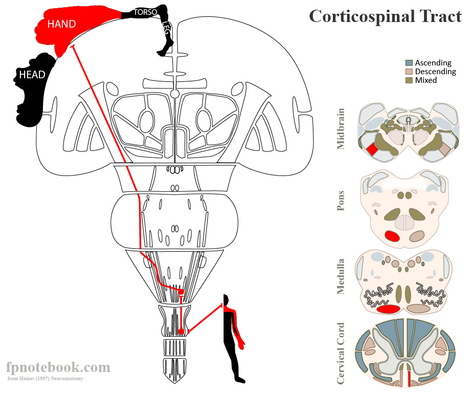
- Corticospinal tract (motor, lateral cord)
- Transmits motor signals from the cerebral cortex
- Pure Corticospinal tract lesions manifest as difficulty with skill movements of the distal extremities
- Most motor deficits (e.g. CVA, posturing) involve mixed pathways
- Fibers cross the midline at the Brain Stem and course to the contralateral Muscles
- Motor fibers Synapse in the anterior horn (grey matter of the spinal cord)
- Upper Motor Neurons (UMN) are from the cerebral cortex to the Synapse
- Lower Motor Neurons (LMN) are from the Synapse to the Muscle
- Images
- Transmits motor signals from the cerebral cortex
- Corticobulbar Tract
- Transmits motor signals from the cerebral cortex to the head
- Extrapyramidal Motor Tracts
- Tectospinal Tract
- Reticulospinal Tract
- Vestibulospinal Tract
- Rubrospinal Tract
VII. Components: Peripheral Nerves
- Sensory
- Enters the spinal cord via the posterior root (at the posterior horn of the grey matter)
- Includes a sensory Ganglion along the posterior root
- Motor
- Exits the spinal cord via the anterior root (at the anterior horn of the grey matter)
VIII. Components: Circulation
-
Anterior Spinal Artery (1)
- Supplies the anterior two thirds of the spinal column
- Arises inferior to the Vertebrobasilar Junction from the 2 Vertebral arteries
- Posterior spinal arteries (2)
- Supplies the posterior one third of the spinal column (in combination with intercostal arteries)
- Arises variably from the Vertebral arteries or posterior inferior cerebellar arteries
IX. Associated Conditions
-
Amyotrophic Lateral Sclerosis (Lou Gehrig's Disease)
- Anterior horns of grey matter (Lower Motor Neuron Deficit)
- Corticospinal tracts (upprer motor Neuron deficit)
-
Tertiary Syphilis (Tabes Dorsalis)
- Posterior Columns and posterior roots and ganglia (proprioception loss and pain)
-
Pernicious Anemia (Vitamin B12 Deficiency)
- Posterior Columns (proprioception loss)
- Corticospinal tracts (Upper Motor Neuron Deficit)
-
Polio
- Anterior horn cells of grey matter (Lower Motor Neuron Deficit) causes weakness, Fasciculations, hyporeflexia
-
Guillain-Barre Syndrome
- Peripheral Nerve deficit (motor and sensory)
-
Syringomyelia
- Idiopathic central spinal cord (or Brain Stem) degeneration
- Affects crossing pain-TemperatureSensation fibers (Spinothalamic Tract)
- Variably affects other structures (e.g. grey matter anterior horn or posterior horn cells)
-
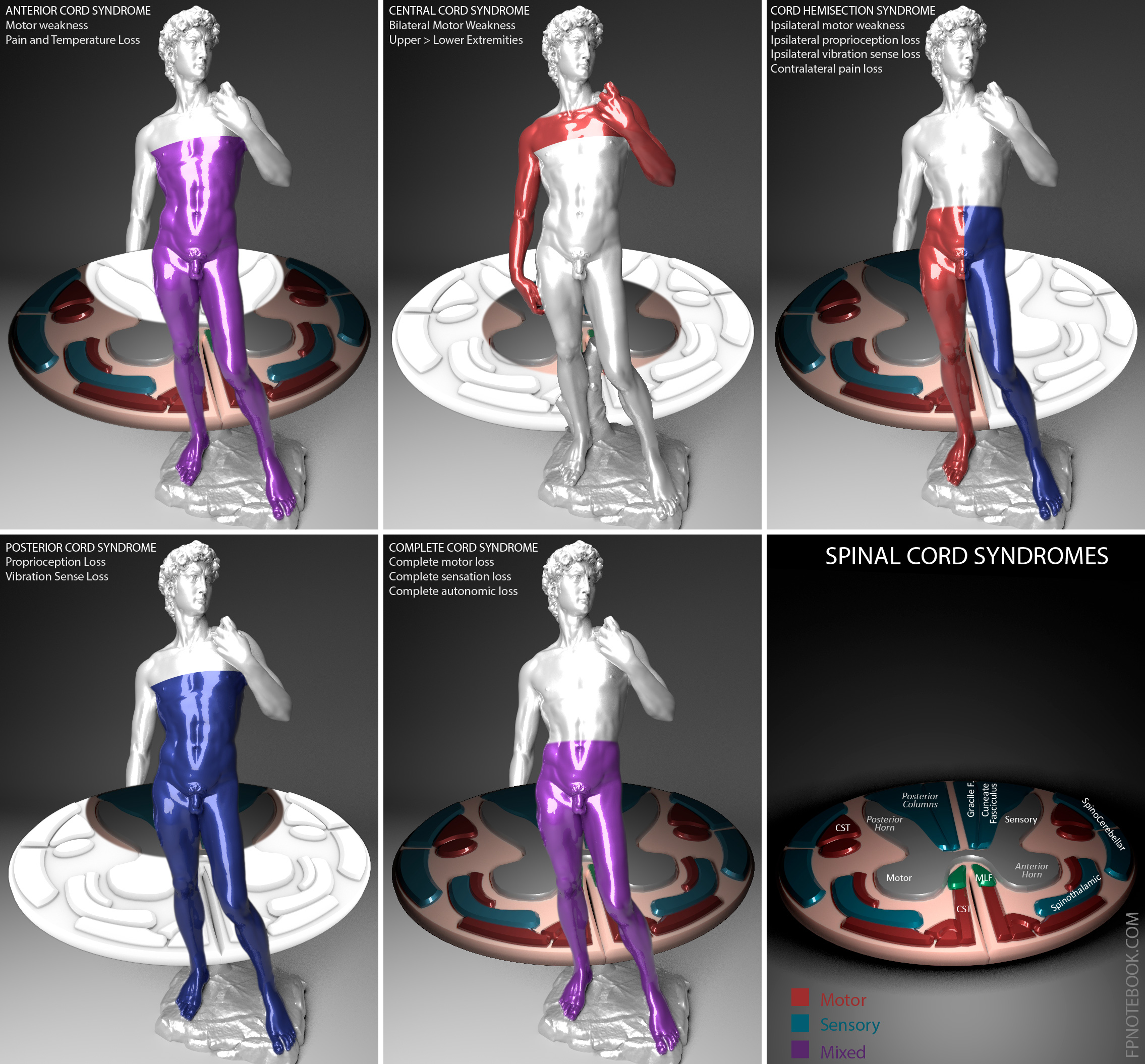
Spinal Cord Syndrome (Trauma)
- Complete transection of the spinal cord (Complete Cord Syndrome)
- Partial spinal cord injuries
- Central Cord Syndrome
- Lesions in the the cervical or thoracic spinal cord
- Result in a relative weakness of the arms in contrast to the legs.
- Anterior Cord Syndrome
- Spinothalamic Tract (pain and Temperature sense)
- Corticospinal tract (motor weakness)
- Posterior Cord Syndrome
- Dorsal Columns (proprioception)
- Spinal Cord Hemisection (Brown-Sequard Syndrome, see below)
- Neuropraxia (Stinger, Burner)
- Central Cord Syndrome
-
Brown-Sequard Syndrome
- Spinal Cord Hemisection
- Results in deficits below the level of the lesion
- Ipsilateral paralysis (Corticospinal tract)
- Ipsilateral proprioception and stereognosis loss (Posterior Columns)
- Contralateral pain and Temperature sense loss (crossed fibers of Spinothalamic Tract)
X. Differential Diagnosis
-
Peripheral Nerve involvement
- Single Dermatome
- Peripheral Neuropathy unique pattern (e.g. Diabetic Neuropathy's stocking-glove distribution)
- Cortical or Brain Stem involvement
- Regional involvement (e.g. an entire extremity sensory or motor)
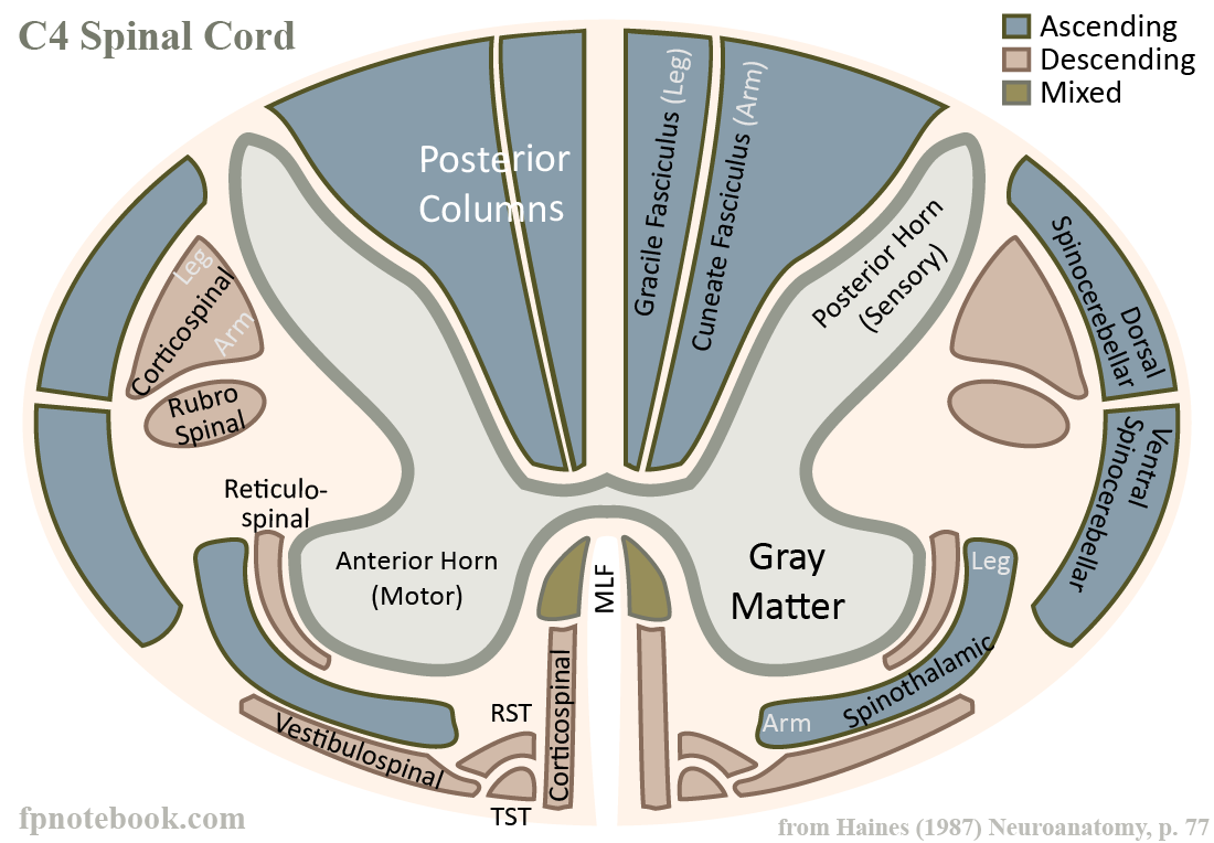
XI. Anatomy: Images
- C4 Level (cross-section)
- Cord Syndromes
- Other images
 Lewis (1918) Gray's Anatomy 20th ed (in public domain at Yahoo or BartleBy)
 Lewis (1918) Gray's Anatomy 20th ed (in public domain at Yahoo or BartleBy)
 Lewis (1918) Gray's Anatomy 20th ed (in public domain at Yahoo or BartleBy)
 Lewis (1918) Gray's Anatomy 20th ed (in public domain at Yahoo or BartleBy)
 Lewis (1918) Gray's Anatomy 20th ed (in public domain at Yahoo or BartleBy)
 Lewis (1918) Gray's Anatomy 20th ed (in public domain at Yahoo or BartleBy)
 Lewis (1918) Gray's Anatomy 20th ed (in public domain at Yahoo or BartleBy)
 Lewis (1918) Gray's Anatomy 20th ed (in public domain at Yahoo or BartleBy)
 Lewis (1918) Gray's Anatomy 20th ed (in public domain at Yahoo or BartleBy)
 Lewis (1918) Gray's Anatomy 20th ed (in public domain at Yahoo or BartleBy)
 Lewis (1918) Gray's Anatomy 20th ed (in public domain at Yahoo or BartleBy)
 Lewis (1918) Gray's Anatomy 20th ed (in public domain at Yahoo or BartleBy)
 Lewis (1918) Gray's Anatomy 20th ed (in public domain at Yahoo or BartleBy)
 Lewis (1918) Gray's Anatomy 20th ed (in public domain at Yahoo or BartleBy)
 Lewis (1918) Gray's Anatomy 20th ed (in public domain at Yahoo or BartleBy)


XII. References
- Gilman (1989) Manter and Gatz Essentials of Neuroanatomy and Neurophysiology, Davis, p. 1-16, 29-37
- Goldberg (2014) Clinical Neuroanatomy, p. 20-3
- Netter (1997) Atlas Human Anatomy, ICON Learning, p. 148-51