
II. Anatomy
- Central CN 5
- Facial Distribution
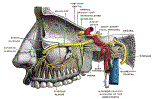
- Trigeminal Nerve
 Lewis (1918) Gray's Anatomy 20th ed (in public domain at Yahoo or BartleBy)
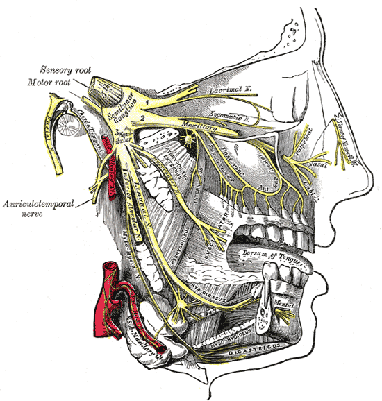
- Alveolar Nerve (V3)
 Lewis (1918) Gray's Anatomy 20th ed (in public domain at Yahoo or BartleBy)


III. Physiology: General
- Largest Cranial Nerve
- Sensory root innervates head and face
- Motor Root innervates mastication Muscles
-
Fibers cross the midline in the Brainstem (at the level of the motor or sensory nucleii)
- Cortical or thalamic CVA involving the trigeminal signals
- Affects the contralateral face
- Brainstem CVA involving the trigeminal signals
- Injures the trigeminal nucleii or crossing fibers, affecting the ipsilateral face
- May injure the Trigeminal Lemniscus affecting the contralateral face
- Cortical or thalamic CVA involving the trigeminal signals
IV. Anatomy
- Trigeminal Lemniscus
- Nucleii (fibers originate in two nuclei)
- Motor Nucleus
- Small and localized to the Pons
- Sensory Nucleus
- Very long nucleus (extends from Midbrain to spinal cord)
- Mesencephalic nucleus of CN 5 (proprioception) is most superior, in the Midbrain
- Main sensory nucleus of CN 5 (light touch) is within the pons
- Spinal nucleus of CN 5 (pain and Temperature sense) is in the lower Medulla and upper spinal cord
- Motor Nucleus
- Course
- Course from pons to trigeminal Ganglion at petrous apex
V. Anatomy: Innervation (Divides into three branches)
- Ophthalmic Branch (Sensory)
- Cornea
- Ciliary body
- Conjunctiva
- Nasal cavity
- Sinuses
- Skin of eyebrows, forehead, and nose
-
Maxillary Branch (Sensory)
- Side of nose
- Lower Eyelid
- Upper lip
- Mandibular Branch (Sensory and Motor)
VI. Exam
- Motor
- Sensory
- Oral Mucosa
- Jaw
- Cheek
- Forehead
- Reflexes
- Jaw Jerk Reflex
- Tests both sensory and motor Trigeminal Nerve
- Corneal Reflex (Cornea touched with cotton whisp)
- Tests both Sensory CN 5 and Motor CN 7
- Consensual reflex results in bilateral blinking
- Jaw Jerk Reflex
VII. Associated Conditions
VIII. References
- Gilman (1989) Manter and Gatz Essentials of Neuroanatomy and Neurophysiology, Davis, p. 87-113
- Goldberg (2014) Clinical Neuroanatomy, p. 24-39
- Netter (1997) Atlas Human Anatomy, ICON Learning, p. 110-129