
II. Definitions
- Neuron
- Specialized conducting cell of the neurologic system which receives, conducts and transmits small electrical signals
- Building blocks of the nervous system
- Neurons are specialized into sensory Neurons and Motor Neurons
- Interneurons are a third type of Neurons that form interconnections between other Neurons
- Multiple Neurons are grouped into pathways
- Peripheral Nervous System: Nerves
- Central Nervous System: Tract, fasciculus, lemniscus, peduncle
- Synapse
- Connections between Neurons in which they communicate via chemical signals (Neurotransmitters)
- Neurologic Pathway
- Chain of communicating Neurons
- Neuraxis Tract (or fasciculus, peduncle or lemniscus)
- Bundle of axons in a pathway within the Central Nervous System (CNS)
- Nerve
- Bundle of axons in a pathway within the Peripheral Nervous System
- Neural Nucleus
- Group of Neuron cell bodies (soma) with attached group of axons (nerve tracts)
- Includes brain nucleii, Cranial Nerve nucleii, cerebellar nucleii and spinal cord nucleii
- Ganglia are the Peripheral Nerve versions of the CNS neural neuclei
III. Anatomy
- Images
- Background
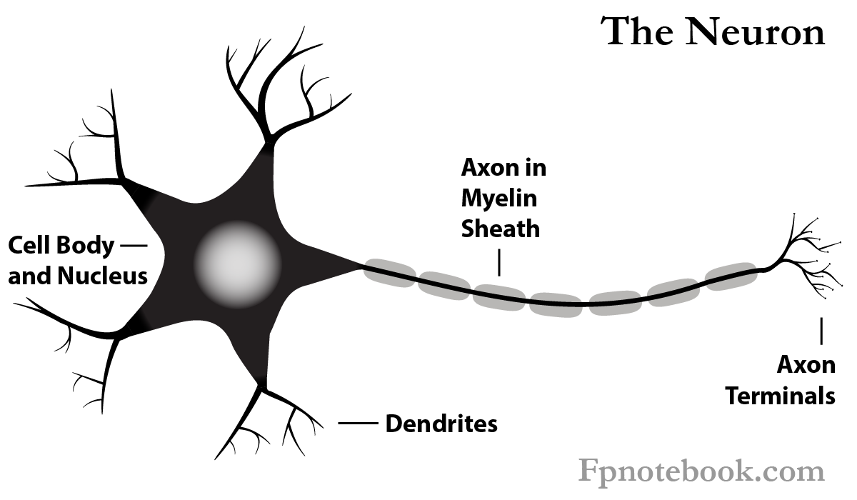
- As with all cells, Neurons have a cell body (soma) with cytoplasm and a nucleus.
- Dendrites
- Tree-like extensions along the cell body that receive signals from other Neurons, or from Sensory Receptors
- Cell Body (Soma)
- Nucleus
- Cytoplasm (Perikaryon)
- Axon
- Transmits signal from cell body to axon terminals
- From the axon terminals, the signal is passed to other nerves via Neurotransmitters across Synapses
- Myelin Sheath
- Most axons are insulated with a thin layer (myelin) of cells to conserve and speed electrical transmission
- Myelinated axons appear as white matter (while Neuron cell bodies appear as gray matter)
- Transmits signal from cell body to axon terminals
-
Neurotransmitters
- Released from axon terminals to transmit a signal into Synapse (inter-Neuron space)
- Neuron types
- Sensory Neurons
- Motor Neurons
- Interneurons (interconnections between Neurons forming a pathway)
- Group of Neurons
- Peripheral Nervous System
- Nerves
- Central Nervous System (interchangeable names)
- Nerve Tract
- Fasciculus
- Lemniscus
- Peduncle
- Peripheral Nervous System
IV. Physiology: Nerve Impulse (Action Potential)
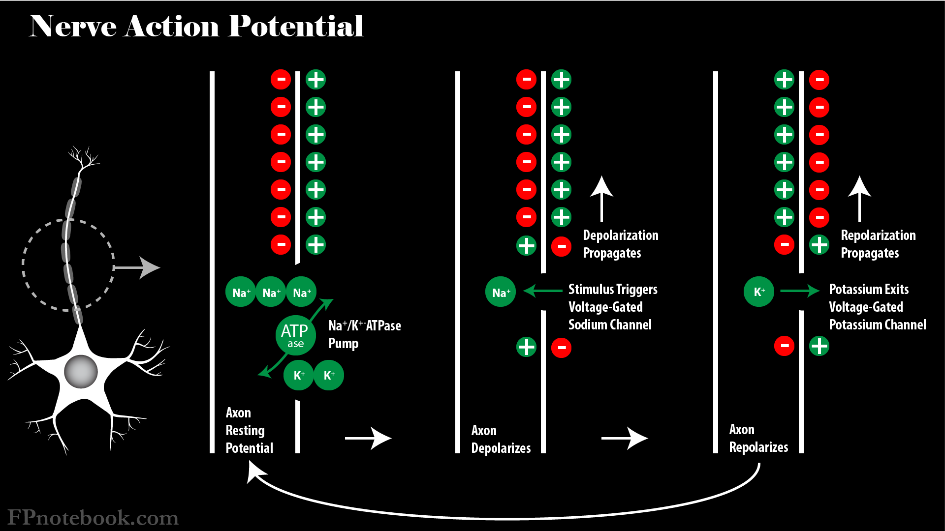
- Neurons are specialized cells capable of tranmsitting an electrical signal
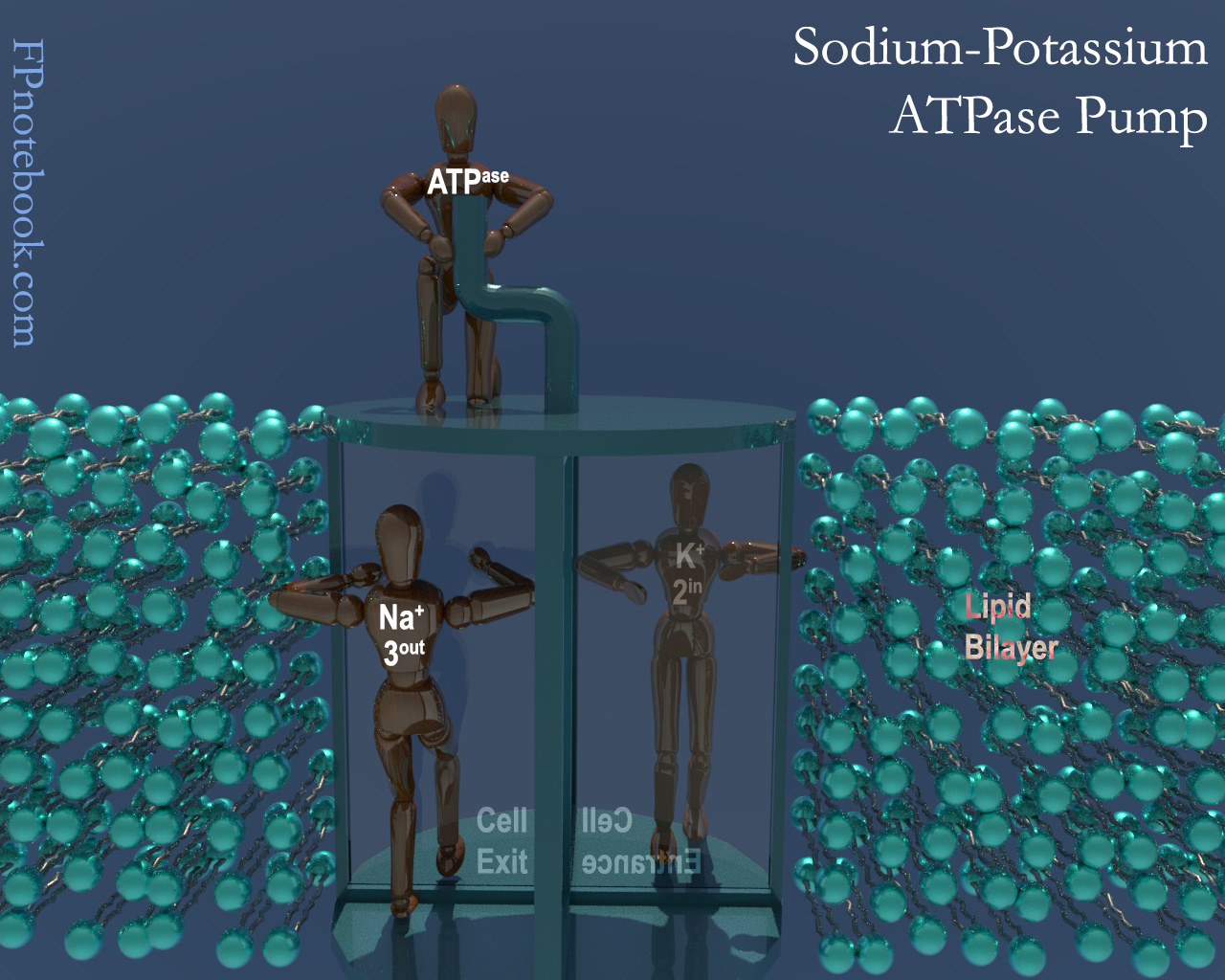
- Neuron resting Membrane Potential is more positive on outside of cell than on inside (e.g. -60 mv difference)
- Depolarization
- Voltage gated electrical channels specific for Potassium and Sodium allow for electrical signal transmission
- Voltage channels are activated when there is a neutralization of resting Membrane Potential
- Membrane Potential decreases below a threshold (e.g. 15-35 millivolts or mv)
- Nerve Depolarization is an all-or-none phenomenon
- Nerve Impulse is only initiated if there is a sufficient Action Potential
- Voltage-Gated Sodium channels suddenly open
- Sodium rushes into Neuron, resulting in neutralization of resting potential and depolarization
- Inside and outside of Neuron may have minimal difference of charge at depolarization
- Voltage-Gated Calcium channels may also be involved
- Most common in cardiac Muscle (esp. Purkinje Fibers) and Smooth Muscle (uncommon in axons)
- As with Sodium, Calcium concentrations outside the cell are higher
- When Calcium channels open, Calcium rushes into the Muscle Cell
- However Calcium channels are slower than Sodium channels
- Results in an Action Potential plateau and a delayed repolarization/recovery
- Allows for a sustained, prolonged contraction of Muscles
- Signal spreads along the axon via contiguous regions, each depolarizing in turn
- Signal amplitude is fixed regardless of the stimulus strength
- However, stronger stimuli result in increased frequency of Action Potential impulses
- Myelin Sheath
- Unmyelinated Peripheral Nerve fibers
- Axon depolarizes continuously, via contiguous ion channels along its surface
- Myelinated Peripheral Nerve fibers
- Axons are insulated, wrapped with surrounding Schwann Cells
- Gaps between the Schwann Cells are known as Nodes of Ranvier
- Ion channels are not exposed where they are covered by overlying Schwann Cells
- Ion channels are only exposed at the Nodes of Ranvier
- Action Potentials must jump between Nodes of Ranvier (Saltatory Conduction)
- Results in most faster depolarization than with unmyelinated fibers
- Axons are insulated, wrapped with surrounding Schwann Cells
- Unmyelinated Peripheral Nerve fibers
- Repolarization
V. Physiology: Synapse
- Synapse is a connection between Neurons in which they communicate via chemical signals (Neurotransmitters)
- Nerve Impulse or Action Potential (see above)
- Nerve Impulse traverses the axon until it reaches the nerve terminals
- Nerve Impulse triggers nerve terminal release of Neurotransmitters from the pre-synaptic membrane
- Neurotransmitters pass into the inter-Neuron space (Synapse)
-
Neurotransmitters
- See Neurotransmitters
- Neurotransmitters act on the post-synaptic membrane of the adjacent Neuron's Dendrites
- Neurotransmitters lower the post-synaptic Membrane Potential of the next Neuron
- Target Neuron Stimulation requires the facilitation of multiple Action Potential triggers to fire
- Stimulation of many Synapses on the same target Neuron (spatial summation) or
- Rapid succession of Action Potentials over relatively few Synapses (temporal summation)
- Each Neuron may have up to 100,000 excitatory and inhibitory inputs that, summed, determine firing potential
- Excitatory Postsynaptic potential (EPSP) refers to sum of excitatory inputs (Action Potentials)
- Inhibitory Postsynaptic potential (IPSP) refers to sum of inhibitory inputs (Action Potentials)
VI. Physiology: Neuronal Networks
- Neurons are interconnected, often with thousands of inputs and outputs at a Synapse
- Neurotransmitters may have excitatory or stimulatory (positive) or inhibitory (negative) effects at the Synapse
- Patterns: Feedback Loops
- Negative Feedback
- Neuron A has excitatory effects at Neuron B
- Neuron A also has excitatory effects at Neuron C
- Neuron C inhibits Neuron A from firing
- Reverbation
- Neuron A has excitatory effects at Neuron B
- Neuron A also has excitatory effects at Neuron C
- Neuron C has excitatory effects at Neuron A, resulting in sustained firing
- Negative Feedback
- Patterns: Inter-Neuron
- Lateral Inhibition
- Neuron A, B and C lie in parallel to one another
- When Neuron B fires, it has excitatory effects on downstream Neurons
- However, Neuron B also has inhibitory effects on Neurons A and C
- Convergence
- Multiple Neurons input to a single Neuron output
- Divergence
- One Neuron has multiple Neuron outputs
- Neural Net
- Multiple Neurons interconnected with one another
- Lateral Inhibition
VII. Pathophysiology: Neurotransmission Disorders
- See Neurotransmitter
- Hyperexcitable Neurons with increased automaticity
- Excessive Neurotransmitter release and activity at post-synaptic receptors
- Insufficient Neurotransmitter release and activity at post-synaptic receptors
VIII. Images
-
 Lewis (1918) Gray's Anatomy 20th ed (in public domain at Yahoo or BartleBy)
-
 Lewis (1918) Gray's Anatomy 20th ed (in public domain at Yahoo or BartleBy)
-
 Lewis (1918) Gray's Anatomy 20th ed (in public domain at Yahoo or BartleBy)
IX. References
- Goldberg (2014) Clinical Physiology, MedMaster, p. 36-7, 87-9