
II. Definitions
- Parietal Lobe
- Parietal Lobe receives sensory input and performs language processing
- As with motor centers in the Frontal Lobe, the Parietal Lobe is organized in the form of the cortical humunculus
- Disproportionately large region devoted to the face and hands, in contrast with the torso and legs
- Sensory functionality is primarily contained in Brodmann Areas 3,1,2
- Also in adjacent seconday somatic area (for pain and TemperatureSensation)
- Brain Lesions result in Receptive Aphasia, sensory loss, Hemianopia and and spatial Disorientation.
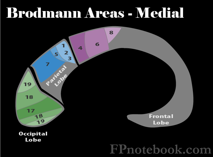
III. Anatomy: Brodmann Areas of Parietal Lobe
- Images
- Primary Somatosensory Cortex or Somesthetic Area (Area 3,1,2)
- Lesions in this region affect contralateral sensory loss in light touch, pressure and proprioception
- Pain and Temperature sense are received in a region inferior to Area 3,1,2, known as the Secondary Somesthetic Area.
- Secondary Somatosensory Cortex or Somesthetic Area (Area S2)
- Lesions in this region affect pain and Temperature sense
- These fibers are inferior to Primary Somesthetic Area (Area 3,1,2)
- Superior Parietal Lobule (Anterior, Area 5)
- Involved in spatial orientation
- Visuo-Motor Coordination (Posterior, Area 7)
- Involved in spatial orientation
- Angular Gyrus (Area 39)
- When in the dominant hemisphere, a lesion in the Angular Gyrus affects the ability to read (alexia) and write (agraphia).
- Supramarginal Gyrus (Area 40)

IV. Physiology: Homunculus
- Homunculus is a graphical representation of sensory and motor control
- Man standing at the midline between the two Cerebral Hemispheres
- Legs are above the falx cerebri at the medial Parietal Lobe (Anterior Cerebral Artery)
- Legs are relatively small compared to arms and head
- Represents fewer overall Neurons dedicated to leg motor and sensory control
- Man is bending at the waist over the top of the Parietal Lobe
- Legs and head extend over the lateral Parietal Lobe (Middle Cerebral Artery)
- Arms and head are relatively large
- Represents greater overall Neurons dedicated to arm/head motor and sensory control
V. Signs: Brain Lesions
- Receptive Aphasia (Area 39)
- Sensory loss (Area 3,1,2 and Area S2 following Homunculus distribution)
- Spatial Disorientation (Area 5, 7)
- Hemianopia (loss of half of Visual Field in each eye)
- Agnosia of tactile Sensation and proprioception (Area 40, dominant hemisphere)
- Apraxia (difficulty with skilled movement) and altered left-right discrimination (Area 40)
VI. Exam
- Cognitive Dominant
- Names fingers
- Knows left and right
- Performs calculations on paper
- Reading
- Cognitive Non-Dominant
- Constructs copy of matchstick figure made by examiner