
II. Definitions
- Temporal Lobe
- Key to longterm visual memories, as well as processing visual and auditory (including language) input
- Supports spontaneous speech, repetition, comprehension, writing and naming
- Lesions may result in behavior change and Memory Loss
- Wernicke's Area (area 22, in the dominant hemisphere) is key to understanding speech and language
- Optic Radiations from the superior Visual Fields also pass through the Temporal Lobes (Quadrantanopia)
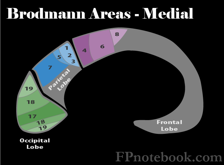
III. Anatomy: Brodmann Areas of Temporal Lobe
- Images
- Wernicke's Area (Area 22)
- Dominant hemisphere lesions in Wernicke's Area result in Receptive Aphasia (auditory Aphasia)
- The patient does not understand speech (including their own speech)
- Auditory Area (Area 41, 42)
- Hearing typically has bilateral input and therefore a lesion affecting one side only may result in no deficit
- Hearing Deficits are typically distal to the exit of Cranial Nerve 8.

IV. History: Findings of pathology (e.g. Tumor)
- Behavior change
- Autistic symptoms
- Memory Loss
V. Exam: Dominant or Standard Aphasia Testing (normal findings)
- Spontaneous speech
- Repetition
- Comprehension
- Writing
- Naming
VI. Exam: Non-Dominant or Affect Interpretation (normal findings)
- Patient names affect in photo of faces or conveyed in examiner's voice
VII. Signs: Brain Lesions
- Complex Partial Seizures or Generalized Seizures
- Quadrantanopia (Vision Loss or anopia in a Visual Field quadrant)
- Behavioral alterations
- Receptive Aphasia (dominant hemisphere, Area 22)