
II. Definitions
- Cerebrum (Telencephalon)
- Largest portion of the Central Nervous System, containing ~15 billion Neurons
- Cerebrum contains the cerebral cortex divided into two hemispheres interconnected by the corpus callosum
- Most people, even those who are left handed, have a dominant left hemisphere.
- Asymmetric higher level functions (e.g. language) are controlled only by the dominant hemisphere
- Each Cerebral Hemisphere is further divided into lobes (Frontal Lobe, Parietal Lobe, Occipital Lobe and Temporal Lobe)
- The Cerebrum also includes the subcortical structures ( Hippocampus, Basal Ganglia, Amygdala, Olfactory Bulb)
III. Background
- Many pathways, such as movement and Sensation, speech and language, Vision and Hearing are well defined.
- Many cerebral regions have been well mapped histologically (e.g. Brodmann Areas) for more than a century
- Modern imaging such as functional MRI have further defined these pathways and regions
- Neurologic roots of personality, emotion, behavior, pleasure are only roughly mapped
- Two Cerebral Hemispheres are each divided into 4 lobes: frontal, parietal, temporal and occipital.
- Symmetric functions (e.g. movement, Sensation)
- Each hemisphere sends and receives signals to the opposite side of the body.
- Asymmetric cerebral functions (e.g. speech and language)
- Left hemisphere is dominant in 95% of right handed and 70% of left handed patients
- Localized to the dominant hemisphere
- Symmetric functions (e.g. movement, Sensation)
IV. Anatomy
- Consistent anatomical landmarks (divide the cerebral lobes into further identifiable regions and maps)
- Brain Gyri (convolutions or ridges)
- Brain sulci and Brain Fissures (involutions or depressions)
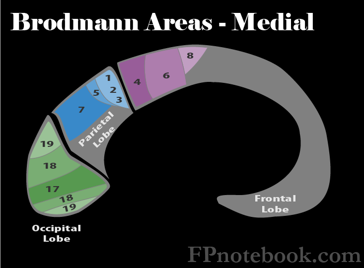
- Brodmann Areas
- Established brain regions by histology in the early 1900s by German researcher (Brodmann)
- See Brodmann Areas of Frontal Lobe
- See Brodmann Areas of Parietal Lobe
- See Brodmann Areas of Temporal Lobe
- See Brodmann Areas of Occipital Lobe
- Brain Lobes
- Frontal Lobe
- Anterior to the central sulcus of Rolando (where the Frontal Lobe meets the Parietal Lobe)
- Function includes personality and verbal expression, as well as motor activity in its posterior aspect
- Parietal Lobe
- Inputs for sensory information including the understanding of speech
- Motor and sensory regions are organized in the form a distorted human (Cortical Homunculus)
- Disproportionately represented large head and hands
- Motor (posterior Frontal Lobe)
- Sensory (anterior Parietal Lobe)
- Occipital Lobe
- Occipital Lobe primarily processes visual input
- Temporal Lobe
- Inferior to the Lateral Fissue of Sylvia (where the Temporal Lobe meets the Frontal Lobe and Parietal Lobe)
- Temporal Lobe relates to behavior and memory
- Frontal Lobe
- Internal Capsule
- Bundles of axons coursing between the cerebral cortex and the Brainstem (one for each hemisphere)
- Composed of three parts: Anterior limb, posterior limb, and genu
- Vascular supply via the Anterior Circulation
- Supplied by small vessels (anterior Choroidal artery, medial striate artery, lateral striate artery)
- Hemorrhage may occur in the supplying vessels with Severe Hypertension
- CVA in this region (despite small distribution) may result in significant neurologic deficits
- Subcortical Region (Subcortex, Diencephalon)

V. Anatomy: Images
- Broad overview - Lobes
 Lewis (1918) Gray's Anatomy 20th ed (in public domain at Yahoo or BartleBy)
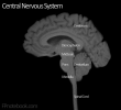
- Miscellaneous
 Lewis (1918) Gray's Anatomy 20th ed (in public domain at Yahoo or BartleBy)
 Lewis (1918) Gray's Anatomy 20th ed (in public domain at Yahoo or BartleBy)
 Lewis (1918) Gray's Anatomy 20th ed (in public domain at Yahoo or BartleBy)
 Lewis (1918) Gray's Anatomy 20th ed (in public domain at Yahoo or BartleBy)
 Lewis (1918) Gray's Anatomy 20th ed (in public domain at Yahoo or BartleBy)
 Lewis (1918) Gray's Anatomy 20th ed (in public domain at Yahoo or BartleBy)
 Lewis (1918) Gray's Anatomy 20th ed (in public domain at Yahoo or BartleBy)
 Lewis (1918) Gray's Anatomy 20th ed (in public domain at Yahoo or BartleBy)
 Lewis (1918) Gray's Anatomy 20th ed (in public domain at Yahoo or BartleBy)
 Lewis (1918) Gray's Anatomy 20th ed (in public domain at Yahoo or BartleBy)
 Lewis (1918) Gray's Anatomy 20th ed (in public domain at Yahoo or BartleBy)
 Lewis (1918) Gray's Anatomy 20th ed (in public domain at Yahoo or BartleBy)
 Lewis (1918) Gray's Anatomy 20th ed (in public domain at Yahoo or BartleBy)
 Lewis (1918) Gray's Anatomy 20th ed (in public domain at Yahoo or BartleBy)
 Lewis (1918) Gray's Anatomy 20th ed (in public domain at Yahoo or BartleBy)
 Lewis (1918) Gray's Anatomy 20th ed (in public domain at Yahoo or BartleBy)
 Lewis (1918) Gray's Anatomy 20th ed (in public domain at Yahoo or BartleBy)
 Lewis (1918) Gray's Anatomy 20th ed (in public domain at Yahoo or BartleBy)
 Lewis (1918) Gray's Anatomy 20th ed (in public domain at Yahoo or BartleBy)
 Lewis (1918) Gray's Anatomy 20th ed (in public domain at Yahoo or BartleBy)
 Lewis (1918) Gray's Anatomy 20th ed (in public domain at Yahoo or BartleBy)
 Lewis (1918) Gray's Anatomy 20th ed (in public domain at Yahoo or BartleBy)
 Lewis (1918) Gray's Anatomy 20th ed (in public domain at Yahoo or BartleBy)
 Lewis (1918) Gray's Anatomy 20th ed (in public domain at Yahoo or BartleBy)
 Lewis (1918) Gray's Anatomy 20th ed (in public domain at Yahoo or BartleBy)


VI. Associated Conditions: Cerebrovascular Accident
VII. References
- Netter (1997) Atlas Human Anatomy, ICON Learning, p. 99-101