
II. Anatomy
-
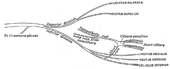
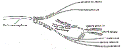 Lewis (1918) Gray's Anatomy 20th ed (in public domain at Yahoo or BartleBy)
-
-

III. Physiology
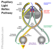
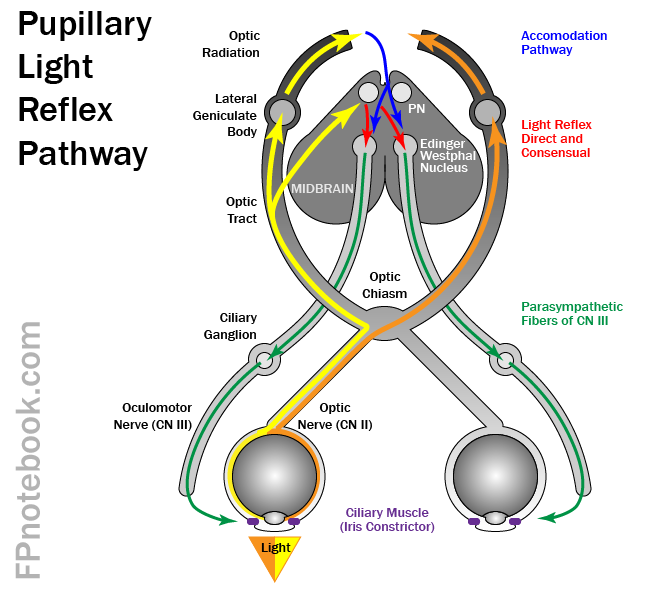
- Parasympathetic fibers follow Cranial Nerve III
- Innervates the ciliary Ganglion which in turn supplies sphincter pupillae and ciliaris Muscles
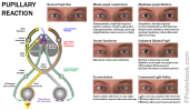
- Parasympathetic impulses result in Pupil Constriction
- Uncal Herniation (Temporal LobeHerniation) compresses the third nerve
- Parasympathetic fibers are most susceptible to injury as they lie on the outside of CN III
- Parasympathetic fiber injury results in an ipsilateral pupil that is fixed/unresponsive and dilated
- With increasing pressure on Cranial Nerve III, complete oculomotor paralysis occurs
- Ultimately contralateral Cranial Nerve 3 involvement ensues
- Innervates five extrinsic eye Muscles
- Levator palpebrae superioris
- Medial rectus
- Superior rectus
- Inferior rectus
- Inferior oblique
IV. Course
- Images
- Nucleii
- Nucleii are located in the floor of Cerebral Aqueduct at the level of the Midbrain
- Oculomotor Nucleus
- Edinger-Westphal Nucleus
- Origin of the visceral motor fibers that innervate pupillary sphincter and ciliary Muscles
- Slightly medial to the Oculomotor Nucleus
- Course
- As with all other Cranial Nerves (except CN 4), fibers remain ipsilateral (do not cross over)
- Exception: Superior Rectus Muscle is innervated by the contralateral Oculomotor Nucleus
- Nerve courses between Superior Cerebellar Artery and Posterior Cerebral Artery
- Forward and lateral to Posterior Clinoid process
- Cavernous Sinus lateral wall
- Enters orbit via superior orbital fissure
- As with all other Cranial Nerves (except CN 4), fibers remain ipsilateral (do not cross over)

V. Exam
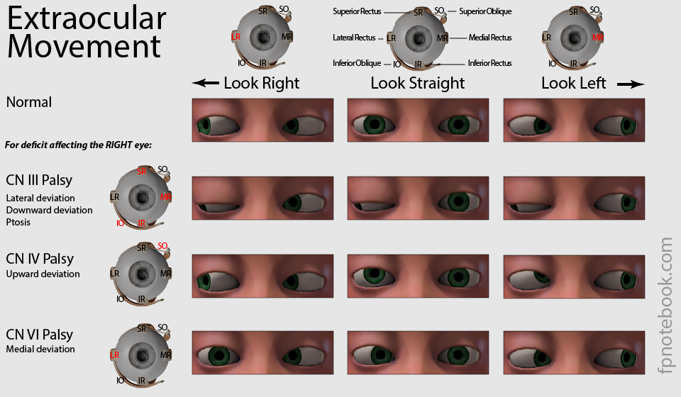
- Extraocular Movement
- CN III Palsy Findings

- Eye Deviation inferolaterally (eye is "down and out")
- Eyelid Ptosis
- Unilateral Pupil Paralysis (Blown Pupil or fixed, non-reactive dilated pupil)
- Oculomotor Nerve Palsy with pupil involvement
- May be due to expanding Cerebral Aneurysm (esp. Posterior Communicating Artery)
- Oculomotor Nerve Palsy with pupil sparing
- Seen as complication with Diabetes Mellitus
VI. References
- Gilman (1989) Manter and Gatz Essentials of Neuroanatomy and Neurophysiology, Davis, p. 87-113
- Goldberg (2014) Clinical Neuroanatomy, p. 24-39
- Netter (1997) Atlas Human Anatomy, ICON Learning, p. 110-129