
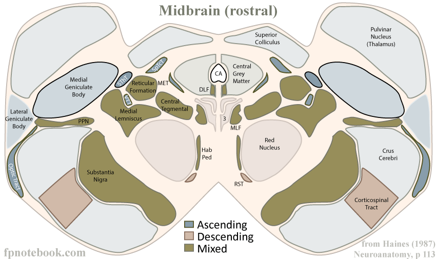
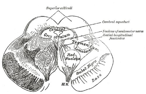
II. Anatomy: Superior (Rostral) Midbrain Components
- Superior colliculus
- Superior (rostral) bump on each side of the upper Midbrain
- Visual reflex center (visual function, orientation of head and eyes)
- Aqueduct
- Single small midline CSF conduit
-
CN 3 nucleus (Oculomotor Nerve)
- Medial, immediately anterior to aqueduct
- Cranial Nerves course anteriorly, and exit in the superior Midbrain at the midline
- Medial Geniculate Body
- Communication relay between the inferior colliculus and the auditory cortex
-
Spinothalamic Tract
- Continued pain and TemperatureSensation tracts from the spinal cord
-
Medial Lemniscus
- Proprioception Sensation from Posterior Columns (via Internal Arcuate Tract)
- Within the tract, head is represented anteriorly and legs posteriorly (as if standing on head)
- Red Nucleus
- Reflexive movement (e.g. crawling)
-
Substantia Nigra
- Substantia Nigra's pars compacta produces Dopamine which is transmitted to the Dorsal Striatum
- Parkinsonism may result if deficient Dopamine production
-
Corticospinal tract
- Continued motor tracts to the spinal cord
- Head is represented anteromedially and legs posterolaterally
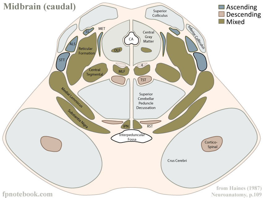
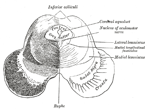
III. Anatomy: Inferior (Caudal) Midbrain Components
- Inferior colliculus
- Inferior (caudal) bump on each side of the lower Midbrain
- Aqueduct
- Single small midline CSF conduit
-
CN 4 nucleus (Trochlear Nerve)
- Medial, immediately anterior to aqueduct
- Cranial Nerves exit posteriorly and immediately cross the midline (unique to CN 4)
-
Spinothalamic Tract
- Continued pain and TemperatureSensation tracts from the spinal cord
-
Medial Lemniscus
- Proprioception Sensation from Posterior Columns (via Internal Arcuate Tract)
- Crossings (Decussation)
- Superior Cerebellar Peduncles Decussation
-
Substantia Nigra
- Substantia Nigra's pars compacta produces Dopamine which is transmitted to the Dorsal Striatum
- Parkinsonism may result if deficient Dopamine production
-
Corticospinal tract
- Continued motor tracts to the spinal cord
IV. Anatomy: Images
- Rostral (superior cross-section)
- Caudal (inferior cross-section)
- Other cross sections
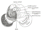 Lewis (1918) Gray's Anatomy 20th ed (in public domain at Yahoo or BartleBy)
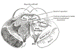 Lewis (1918) Gray's Anatomy 20th ed (in public domain at Yahoo or BartleBy)
V. References
- Gilman (1989) Manter and Gatz Essentials of Neuroanatomy and Neurophysiology, Davis, p. 81-96
- Goldberg (2014) Clinical Neuroanatomy, Medmaster, p. 24-39
- Netter (1997) Atlas Human Anatomy, ICON Learning, p. 108-12