
II. Definitions
III. Background
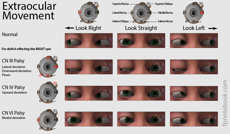
- Three different nerves rotate the eye's axis (line of sight)
-
Oculomotor Nerve (CN 3) innervates 3 of the 5 eye Muscles
- Allows the eye to look up, down and medially
- CN 3 Palsy results in the eye is oriented 'down and out' (functionality of the 2 remaining nerves)
-
Trochlear Nerve (CN 4) innervates the Superior Oblique Muscle
- Pulley system (trochlea) to rotate the eye downward and laterally
-
Abducens Nerve (CN 6) innervates the Lateral Rectus Muscle
- Rotates the eye laterally
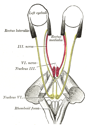
- Conjugate Gaze (alignment of both eyes) is the most complex Extraocular Movement
- Requires one eye to look laterally (CN 6) while the other eye looks medially (CN 3)
- Synchrony requires coordination from several centers
- Contralateral cerebral cortex (Brodmann Areas 17,18,19 and 8)
- Lateral Gaze Center (Pontine Paramedian Reticular Formation or PPRF)
- Signals to ipsilateral CN 6
- Signals to contralateral CN 3 (via Medial Longitudinal Fasciculus)
IV. Anatomy: Innervation
- Cerebral Cortex
- Voluntary Conjugate Gaze (Brodmann's Area 8)
- Involuntary Conjugate Gaze (Areas 17-19)
- Cranial Nerves
- Nucleii and Pathways
- Lateral Gaze Center (Pontine Paramedian Reticular Formation, Paraabducens Nucleus)
- Responsible for horizontal gaze and saccade eye movements
- Facilitates Conjugate Gaze by synapsing with contralateral fibers for coordinated eye movements
- Medial Longitudinal Fasciculus (MLF)
- Transmits signals between CN 3 and CN 6 to coordinate eye movements (Conjugate Gaze)
- Dysfunctional in Internuclear Ophthalmoplegia (e.g. Multiple Sclerosis, esp. if bilateral)
- Example of normal Conjugate Gaze pathway via Medial Longitudinal Fasciculus
- Cortical signal from voluntary gaze center directs left eye to look left
- Left eye CN 6 (lateral rectus) fires for the left eye to look left
- CN 6 signals Synapse with the Lateral Gaze Center
- Signals are transmitted via Medial Longitudinal Fasciculus across midline to contralateral CN 3
- Medial rectus directs the right eye to also look left (adduct to look medially)
- Lateral Gaze Center (Pontine Paramedian Reticular Formation, Paraabducens Nucleus)
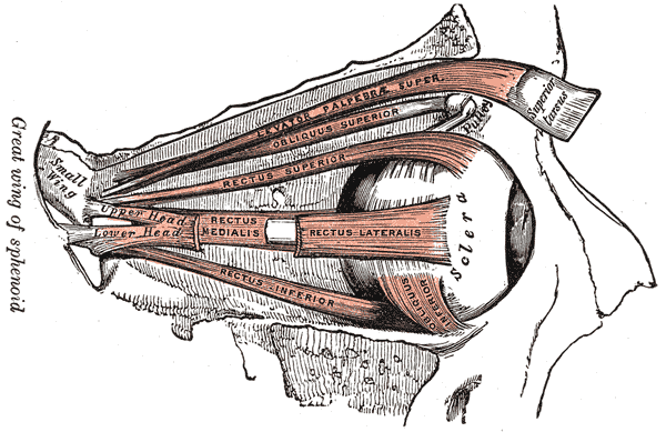
V. Anatomy: Extraocular Muscles (Innervated by CN 3 with 2 exceptions)
- See Brainstem CVA
- Background
- Medial Range of Motion
- Medial Rectus Muscle (Cranial Nerve 3)
- Lateral Range of Motion
- Lateral Rectus Muscle (Cranial Nerve 6)
- Upward Range of Motion
- Superior Rectus Muscle (Cranial Nerve 3)
- Inferior Oblique Muscle (Cranial Nerve 3)
- Downward Range of Motion
- Inferior Rectus Muscle (Cranial Nerve 3)
- Superior Oblique Muscle (Cranial Nerve 4, Trochlear Nerve)
- Pulley system (trochlea) to rotate the eye downward and laterally
- CN 4 Paralysis results in vertical Diplopia, and Head Tilt compensating for eye rotation
- Function depends on eye position
- Eye looks down (nasal position)
- Eye rotates (temporal position)
- Eye looks down and out (neutral straight ahead position)
- Other extraocular Muscles affected Eyelid position
- Upper Eyelid Opening
- Levator Palpebrae Superioris Muscle (CN 3)
- Defect results in significant Ptosis
- Muller's Muscle or Orbitalis Muscle (cervical Sympathetic Nerves)
- Defect results in mild Ptosis
- Levator Palpebrae Superioris Muscle (CN 3)
- Upper Eyelid Closure
- Orbicularis Oculi Muscle (Cranial Nerve 7, defective in Bell's Palsy)
- Upper Eyelid Opening
VI. Anatomy: Images
- See Neurologic Anatomy of the Eye
-

-
 Lewis (1918) Gray's Anatomy 20th ed (in public domain at Yahoo or BartleBy)
-
 Lewis (1918) Gray's Anatomy 20th ed (in public domain at Yahoo or BartleBy)
VII. Pathophysiology
- See Eye Deviation
VIII. References
- Goldberg (2014) Clinical Neuroanatomy, Medmaster, p. 40-53
- Netter (1997) Atlas Human Anatomy, ICON Learning, p. 114, 126