
II. Anatomy
- See Cervical Spine Anatomy
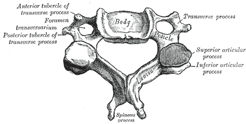
- Image: Cervical Vertebrae
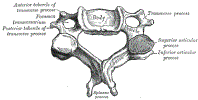 Lewis (1918) Gray's Anatomy 20th ed (in public domain at Yahoo or BartleBy)
- Three-Column Model
- Anterior column
- Anterior half of Vertebral body
- Anterior ligamentous complex
- Anterior portion of annulus fibrosus
- Anterior longitudinal ligament
- Middle column
- Posterior half of Vertebral body
- Ligaments
- Posterior portion of annulus fibrosus
- Posterior longitudinal ligament
- Posterior Column
- Facet joints (superior and inferior articular process)
- Laminae
- Spinous processes
- Posterior ligamentous complex
- Facet capsules
- Interspinous ligaments
- Anterior column
III. Types: C1 Fractures and Dislocations
- Mechanism
- Axial load or Hyperextension injuries
- May occur in diving injury into a shallow pool or being struck on the top of the head by a heavy object
- Jefferson Fracture (C1 Burst Fracture)
- Atlantooccipital Dislocation
IV. Types: C2 Fractures
- Hangman's fracture
- Mechanism
- Previously most associated with hanging (MVA is most common modern mechanism)
- Unstable, hyperextension injury
- Findings
- Mechanism
- Odontoid Fractures (forced flexion or extension)
- Unstable Fracture (esp. types 2-3) secondary to multidirectional injury
- Types (Anderson and D'Alonzo Classification)
- Type 1 (Tip Fracture, most common)
- Avulsion Fracture with injury to the alar ligament
- Fracture of the top of the dens (superior, odontoid tip)
- Instability is uncommon (but evaluate with flexion and extension films)
- Managed with Cervical Collar immobilization
- Type 2 (Waist Fracture, most serious/unstable)
- Fracture at the mid-dens
- High risk for devascularization and nonunion
- Types (Grauer Classification)
- Type 2A: Nondisplaced or minimally displaced and without comminution
- Treated with external immobilization
- Type 2B: Displaced Fracture - anterosuperior to posteroinferior
- Treated with anterior odontoid screw (requires adequate bone density)
- Type 2C: Comminuted, displaced Fracture - anteroinferior to posterosuperior
- Treated with posterior stabilization
- Type 2A: Nondisplaced or minimally displaced and without comminution
- Type 3 (Base Fracture)
- Type 1 (Tip Fracture, most common)
- References
V. Types: Facet Dislocation
- Unilateral facet dislocation
- Bilateral facet dislocation
- Unstable injury
- Mechanism
- Severe hyper-flexion force to the middle and Posterior Columns
- Disrupts the anterior longutudinal ligament, intervertebral disc and posterior ligaments
- Findings
- Types (in order of increasing severity)
- Subluxed facets
- Perched facets
- Locked facets
VI. Types: Wedge Compression Fracture
- Mechanism
- Hyperflexion loading of the spine (even minor forces, esp. in Osteoporosis)
- More often affects the thoracolumbar spine
- Anterior Vertebral body compression (with or without posterior compression)
VII. Types: Flexion Teardrop Fracture
- Mechanism
- Flexion and compression injury (e.g. diving)
- Disrupts all supportive ligaments and the intervertebral disc
- Associated with anterior Spinal Cord Compression (Anterior Cord Syndrome)
- Fracture dislocation which is highly unstable
- References
- Flexion Teardrop Fracture (Radiopaedia)
VIII. Types: Translation-Rotation Fracture
- Severe, unstable injury almost always requiring Spine Surgery
- Mechanism
- Displacement of a Fracture in the horizontal plane (left-right, anterior-posterior or rotational)
- Findings
IX. Types: Burst Fracture
- Mechanism
- Flexion and compression injury (e.g. diving)
- Compression of both the anterior and posterior Vertebral body height
- Most commonly affects the mid-Cervical Spine
- Comminuted, unstable Vertebral Fracture
- Disrupts anterior and middle columns
- Typically involves middle and lower Cervical Vertebrae
- Spinal Cord Injury may result from bone fragments displaced into the spinal canal
- Findings
- Vertebral height loss
- Posterior Ligamentous Complex Injury
X. Types: Spinous Process Fracture
- Mechanisms
- Direct spinous process Trauma
- Sudden deceleration
- High velocity Trauma with neck flexion
- Severe Muscle Contraction with secondary avulsion
- Clay Shoveler's Fracture (Spinous process tip avulsion)
XI. Imaging
XII. Management
- See Cervical Spine Injury
- See Cervical Spine Immobilization
-
Vertebral Fracture Stability
- Consider any cervical Vertebral Fracture unstable with the exception of those listed below
- Subaxial Injury Classification and Severity Scale (SLICS)
- Unstable Cervical Spine Fracture (Mnemonic - "Jefferson Bit Off A Hangman's Tit")
- J - Jefferson Fracture (C1 Burst Fracture, axial loading injury)
- B - Bifacet dislocation or Fracture (flexion injury)
- O - Odontoid Fracture (Types 2 and 3, flexion injury)
- A - Any Fracture-dislocation, Atlantoaxial dislocation or atlanto-occipital dislocation (flexion injury)
- H - Hangman's fracture or bilateral C2 Pedicle Fracture (posterior C2 Fracture, extension injury)
- T - Flexion Teardrop Fracture
- Stable Fractures
XIII. References
- Dreis (2020) Crit Dec Emerg Med 34(7):3-21
- Eiff and Hatch (2018) Fracture Management for Primary Care, p. 187-96
- Ouellette and Tetreault (2015) Clinical Radiology, Medmaster, Miami, p. 42-50