
II. Indications
- Rib Fracture related pain
- Regional Anesthesia for Chest Tube Placement
- Axillary abscess drainage
- Intractable regional pain (e.g. Herpes Zoster)
III. Contraindications
IV. Preparation: Anesthetic
-
Bupivacaine
- Limit Bupivacaine max dose to 2.5 mg/kg or 175 mg for a 70 kg person (risk of LAST Reaction)
- Bupivacaine 0.5% concentration is 5 mg/ml (for a 70 kg person, max of 35 ml Bupivacaine 0.5%)
- Bupivacaine 0.25% concentration is 2.5 mg/ml (for a 70 kg person, max of 70 ml Bupivacaine 0.25%)
- Limit Bupivacaine max dose to 2.5 mg/kg or 175 mg for a 70 kg person (risk of LAST Reaction)
- Dilute in Saline
- Combine Bupivacaine and Normal Saline for a total volume of 30 ml
V. Preparation: Other Materials
- Linear, high frequency Ultrasound probe
- Sterile Ultrasound cover (gel inside of cover) and sterile gel over surface
- Position Ultrasound machine on opposite side of patient
- Needles (choose one)
- Specific Nerve Block needle (magnetized, more easily seen on Ultrasound)
- Blunt tipped spinal needle with IV Line extension tubing
- Syringe (30 ml)
- Second operator to inject Anesthetic
- First operator will hold needle in position
- Skin Preparation
- Sterile Gloves
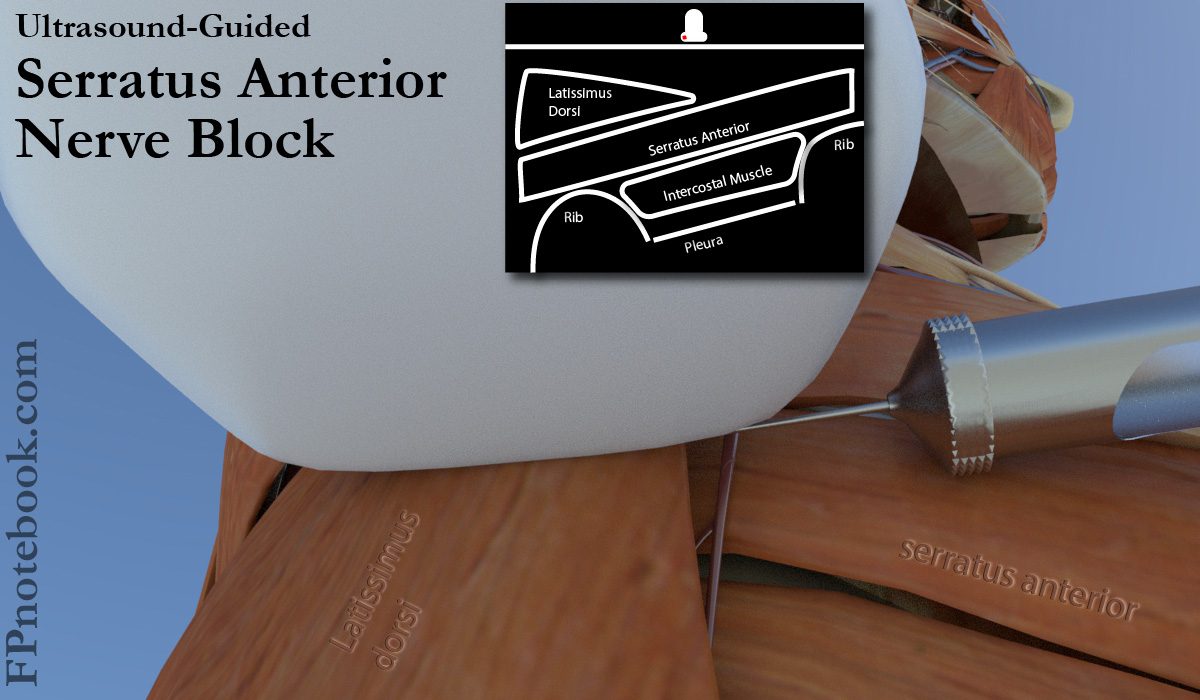
VI. Technique: Ultrasound Guided Serratus Anterior Nerve Block
- Images
- Landmarks: Serratus Anterior insertion site
- Identify rib spaces 4-5 at mid-axillary line (Nipple line in men)
- Ultrasound appearance (from superficial to deep)
- Patient positioning: Lateral decubitus with unaffected side down (preferred)
- Arm on affected side positioned out of the way
- Ultrasound probe placed in horizontal, transverse position over landmark
- Needle directed inline toward table and medial chest
- Patient positioning: Supine
- Arm on affected side raised overhead
- Ultrasound probe placed in horizontal, transverse position over landmark
- Needle directed inline toward lateral chest
- Needle Insertion
- Direct needle toward a rib (acts as a stop in case of patient movement)
- Injection while inserting needle (hydrodissection) identifies the needle tip location on Ultrasound
- Injection Depth
- Injection depth is typically taught to be between Latissimus Dorsi Muscle and Serratus Anterior Muscle
- However injection may be placed either superficial or deep to the serratus anterior
- Superficial surface is preferred to avoid Pneumothorax
- Latissimus dorsi junction is used only as an Ultrasound landmark to identify serratus anterior
- However, serratus anterior is easily identified overlying the ribs in the axilla
- Injection may be anywhere along the serratus anterior surface to provide effective plane block
- Anesthetic Injection
VII. Complications
VIII. Resources
- Ultrasound Guided Serratus Anterior Blocks (Stat Pearls)
- Serratus Plane Block for Rib Fractures (Highland Ultrasound)
IX. References
- (2022) HQMedED Regional Anesthesia for Acute Care Providers, Minneapolis, MN, attended 6/3/2022
- Avila in Herbert (2020) EM:Rap 20(8); 1
- Karsh (2021) CRit Dec Emerg Med 35(3):9