
II. Indications
- Anesthesia of the dorsum of the foot
III. Preparation
- Needle: 27 gauge 1.5 inch
- Skin Preparation (e.g. Hibiclens or Betadine)
-
Anesthetic
- See Regional Anesthesia for Anesthetic options
- Local Anesthetic 2-5 ml (Ultrasound) or 5-10 ml (landmark)
- Bedside Ultrasound (high frequency linear probe) guidance is recommended
IV. Technique: Saphenous Nerve Block
- Indications
- Anesthesia of the medial ankle and medial foot
-
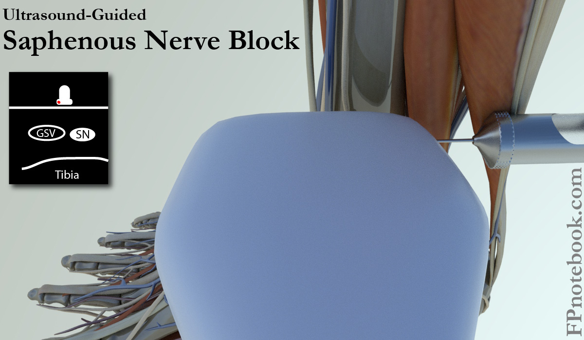
Ultrasound guided (preferred)
- Images
- Patient position
- Lateral decubitus position with the medial ankle upright (or patient supine)
- Ultrasound probe
- Linear probe positioned transverse proximal to the medial malleolus, at distal medial thigh
- Saphenous vein and nerve run between vastus medialis (lateral) and sartorius Muscle (medial)
- Injection
- Insert needle inline with probe from anterolateral to posteromedial
- Saphenous nerve is adjacent to greater saphenous vein (visualize the needle tip!)
- Images
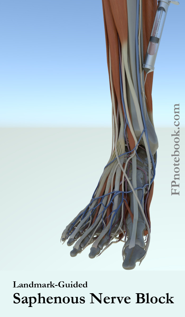
- Landmark Based
- Images
- Patient position
- Supine position with knee flexed and foot rests on table
- Injection
- Inject superior (proximal) and anterior to medial malleolus
- Direct the needle at 45 degree angle posteromedially
- Inject 3 cc subcutaneously along the course (but not within) the greater saphenous vein
- Greater saphenous vein parallels the saphenous nerve
- Images
V. Technique: Deep Peroneal Nerve Block
- Indications
- Anesthesia of the first web space
- Landmarks
- Tibia (medial)
- Extensor Hallucis Longus Tendon
- Deep Peroneal Nerve
- Dorsalis Pedis Artery (midline ankle)
-
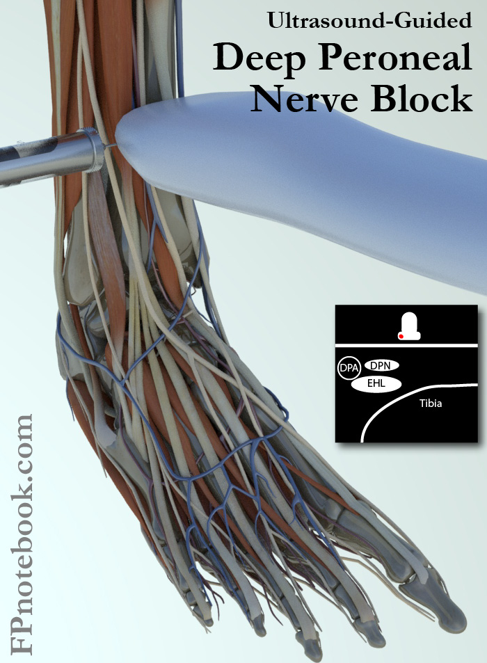
Ultrasound guided (preferred)
- Images
- Position
- Patient Supine
- Ultrasound Probe
- Linear probe transverse to anterior ankle
- Injection
- Inline injection of Anesthetic at the deep peroneal nerve
- Images
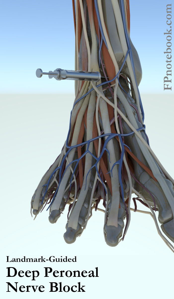
- Landmark Based
- Images
- Position
- Landmarks
- Identify extensor hallucis longus tendon with great toe dorsiflexion against resistance
- Injection site is at level between malleoli, lateral to extensor hallucis longus (and medial to tibialis anterior)
- Injection
- Insert needle toward tibia at site 0.5 cm lateral to extensor hallucis longus tendon
- After striking tibia with needle, withdraw a few mm and aspirate to confirm not within artery
- Inject 5 cc Anesthetic
- Images
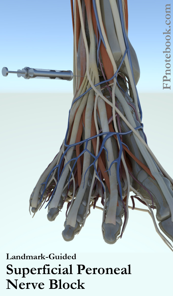
VI. Technique: Superficial peroneal nerve
- Indications
- Anesthesia of most of dorsal foot (and anterolateral leg)
-
Ultrasound-Guided (preferred)
- Images
- Position
- Lateral decubitus position with lateral ankle up
- Ultrasound Probe
- Linear probe transverse to lateral ankle at 3-4 cm superior (proximal) to the lateral malleolus
- Landmarks
- Injection
- Inline injection of Anesthetic at the superficial peroneal nerve
- Images
- Landmark Based
- Images
- Position
- Patient supine with knee flexed and foot resting on table
- Landmarks
- Anterior tibial border AND
- Superior to lateral malleolus
- Injection
- Inject superior to lateral malleolus at 10-15 degree angle towards anterolateral tibia
- Inject 5 cc along the course of the needle
- Images

VII. Precautions
- Avoid injecting directly into nerves
- Distal Paresthesias with needle with injection
- Indicates needle is in nerve
- Do not inject here!
- Remove needle and reposition
VIII. References
- Pfenninger (1994) Procedures, Mosby, p. 1036-54
- Salam (2004) Am Fam Physician 69(4):896 [PubMed]
- Yurgil (2020) Am Fam Physician 101(11):654-64 [PubMed]