
II. Anatomy
-
-
-
-
 Lewis (1918) Gray's Anatomy 20th ed (in public domain at Yahoo or BartleBy)

III. Anatomy: Surrounding landmarks
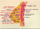
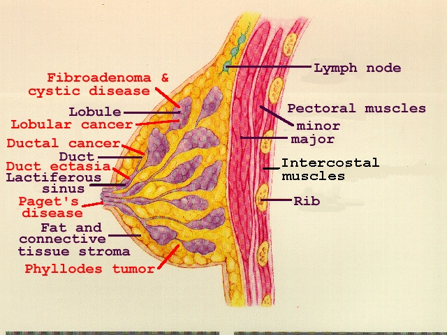
IV. Anatomy: Organization
- Structural Components
- Peri-Nipple structures
- Areola
- Areolar glands
- Nipple
- Cooper's Ligaments
- Add structure to Breast
- Attached to underlying fascia
- Peri-Nipple structures
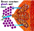
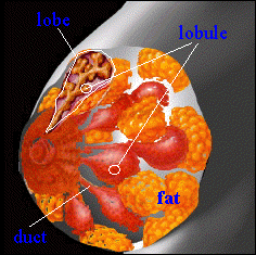
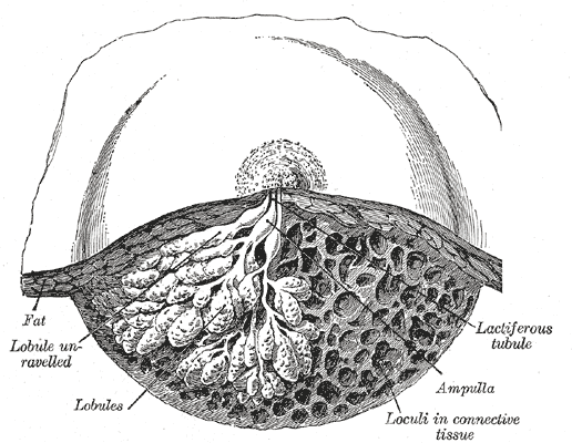
- Key Lactation Components: Ducts and Lobules
- Lobules
- Composed of acini and ducts
- Lobes
- Composed of Lobules
- Lactiferous ducts
- Drain lobes to ducts at nipple surface (6-10 major ducts drain to surface)
- Lobules
V. Physiology: Development
- Pre-Puberty
- Breasts in resting state
- Nonfunctional ducts
-
Puberty: Stage 1 Mammogenesis
- Ovulation starts
- Menstrual Cycle related increase in Estrogen and Progesterone
- Ducts elongate due to Estrogen
- Type 1 Breast lobules develop
- Breast alveolar buds form
- Appear as retroareolar mass (Do not excise as mass! - halts Breast development)
- Type 2 and 3 Breast lobules form from the alveolar buds
- Side branches of ducts and lobular elements form
- Ovulation starts
- Maturity (end of Puberty)
- Further Breast development stops at the end of Puberty (until pregnancy)
- Breasts become pendulous
- Lobular elements well formed in resting state
- Pregnancy
- General Changes
- Areolar pigmentation
- Vascular engorgement
- Mammary Blood Flow increases 180%
- Breast doubles in weight
- May result in bloody Nipple Discharge
- Second and third trimester and early Lactation
- Resolves spontaneously in most cases
- Stage 2 Mammogenesis (first half of pregnancy)
- Breast alveoli develop in response to increased Estrogen and Progesterone levels
- Proximal ducts grow and branch
- Type 3 Breast lobules form in response to HCG in early pregnancy
- Breast lobulesn as well as the overall Breast increase in size during pregnancy
- Stage 1 Lactogenesis or Secretory Initiation (second half of pregnancy)
- Increased Progesterone from the placenta inhibits Prolactin and milk production
- Small amounts of milk and colostrum form
- Stage 2 Lactogenesis or Secretory Activation (after delivery)
- Progesterone levels fall (without placenta)
- Proalctin is no longer inhibited by Progesterone, and Lactation occurs
- Prolactin
- Stimulates copious milk production
- Prolactin secretion is inhibited by Dopamine activity at D2 receptors on the Hypothalamus
- Other Lactation triggers
- Cortisol
- Insulin
- Nipple stimulation
- Emptying of the Breast (by Breast Feeding or pumping)
- Oxytocin
- Stimulates milk let down, myoepithelial cell contraction and milk ejection
- Progesterone levels fall (without placenta)
- General Changes
- Perimenopausal
- Lobules begin to recede
- Leaves residual ducts and fibro-connective tissue
- Breast cysts commonly develop during this stage
- Postmenopausal
- Residual ducts and fat
- Easiest time for Clinical Breast Exam
VI. References
- Pillay (2022) Lactation Physiology, Stat Pearls, Treasure Island, Fl, accessed 2/24/2023