
II. Technique: Transducer
- Transducer orientation (start)
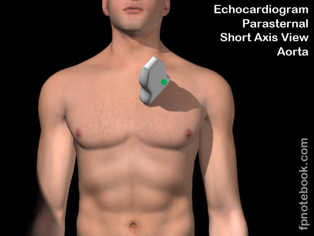
- Transducer Rotated 90 degrees clockwise from Parasternal Long Axis View
- Transducer 3-5 cm to the left of the left sternal border at 3rd to 5th intercostal space
- Transducer indicator pointed towards patient's left Shoulder (1:00 position)
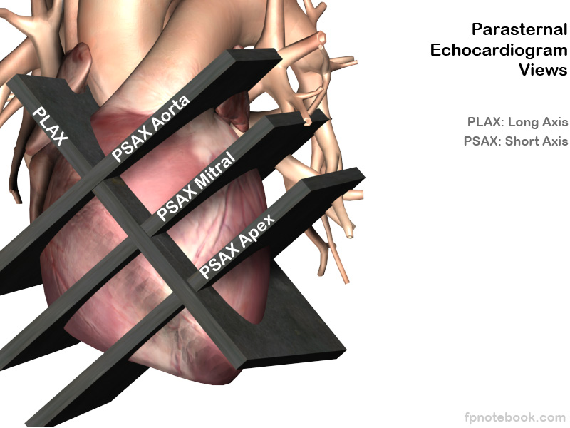
- Images
- Transducer gradually tilted down heart axis to obtain 4 heart cross-sectional slices
- Aortic valve level (may require sliding up a rib space)
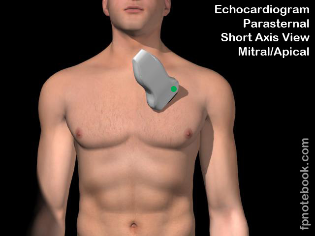
- Mitral valve level
- Mid-ventricle level
- Heart apex (may require sliding down a rib space)
- Aortic valve level (may require sliding up a rib space)
III. Technique: Landmarks
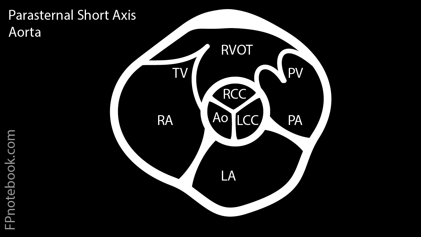
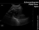
- Landmarks: Aortic valve level
- Right ventricular outflow tract
- Tricuspid valve, aortic valve (peace or mercedes sign when tri-leaflet) and pulmonic valve
- Right atrium, left atrium and pulmonary artery
- Right and left main Coronary Artery origins may be visualized just cephalad to the aortic level
- Color Flow, pulse wave and Continuous Wave Doppler may be used for Tricuspid insufficiency and Pulmonic Insufficiency
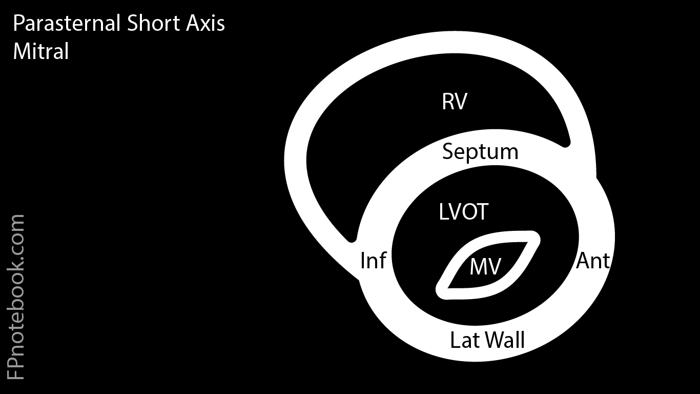
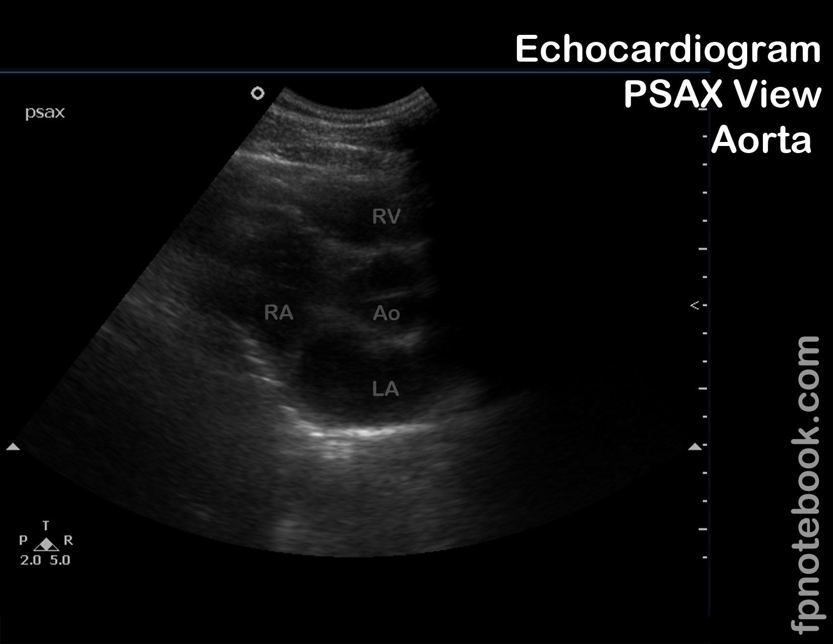
- Landmarks: Mitral valve level
- Right ventricle
- Mitral valve (anterior and posterior leaflets appear as a fish mouth opening and closing)
- Left ventricle should be round (not an oblong oval); rotate the probe until round
- Mitral Valve area may be estimated by planimetry (drawn boundary of open valve)
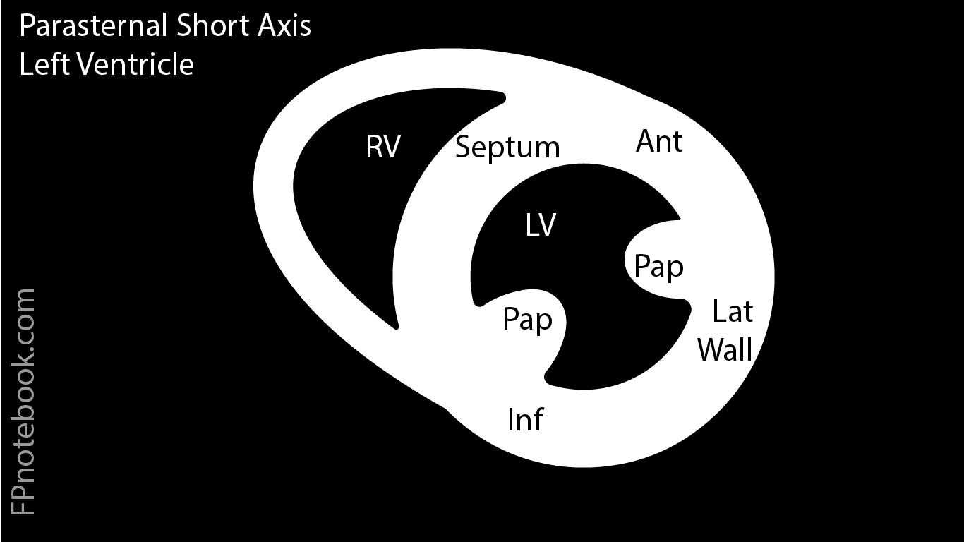
- Landmarks: Mid-ventricle level (most useful short axis view for emergency department)
- Allows for observation of ventricular wall motion and overall contractility, as well as comparison of RV vs LV diameter
- Right ventricle
- Septum
- Left ventricle (with trabeculations representing papillary Muscles)
- Mnemonic: SALI (from Richie Palma, who calls the LV on PSAX, his girlfriend "Sally")
- From the Septum, rotating clockwise: S-Septum, A-Anterior, L-Lateral, I-Inferior
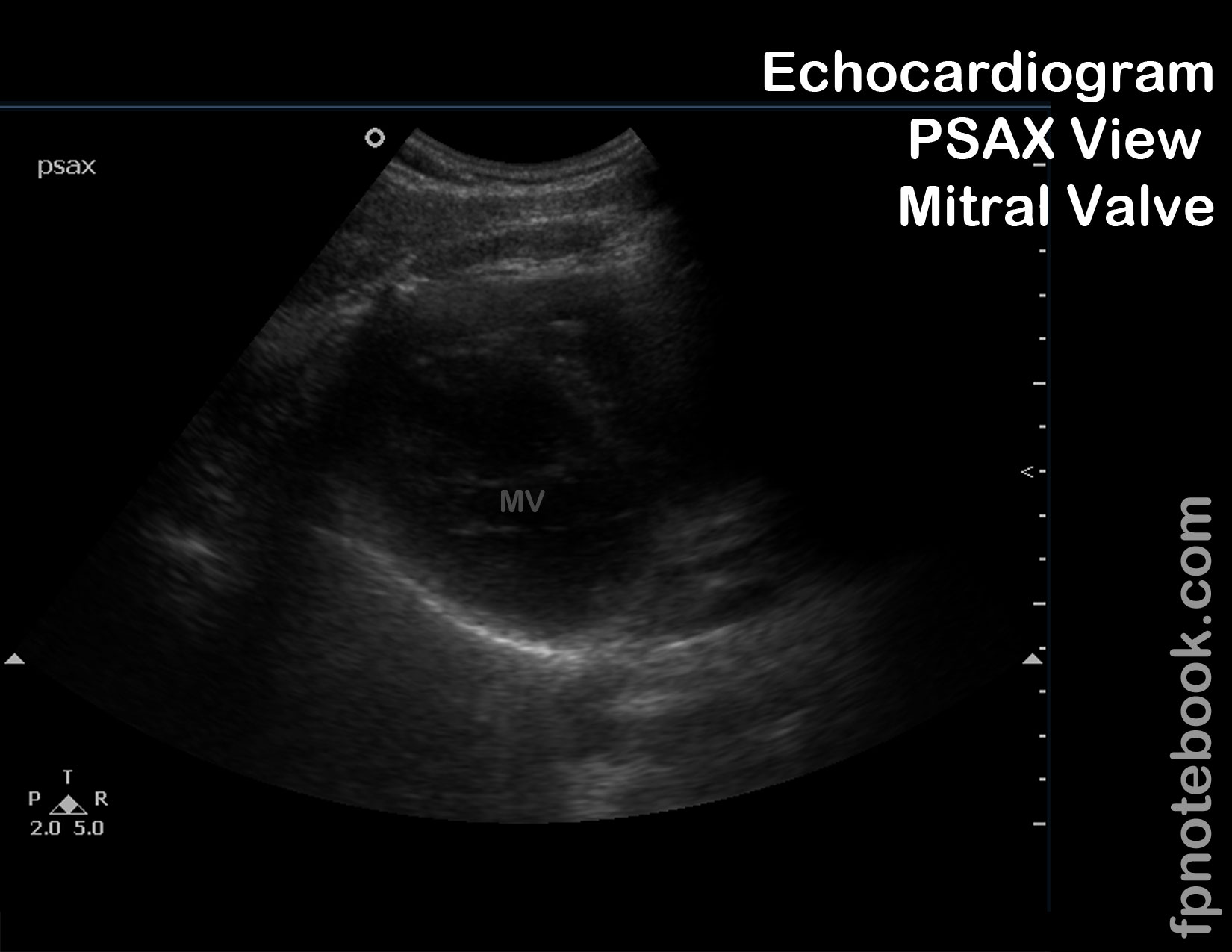
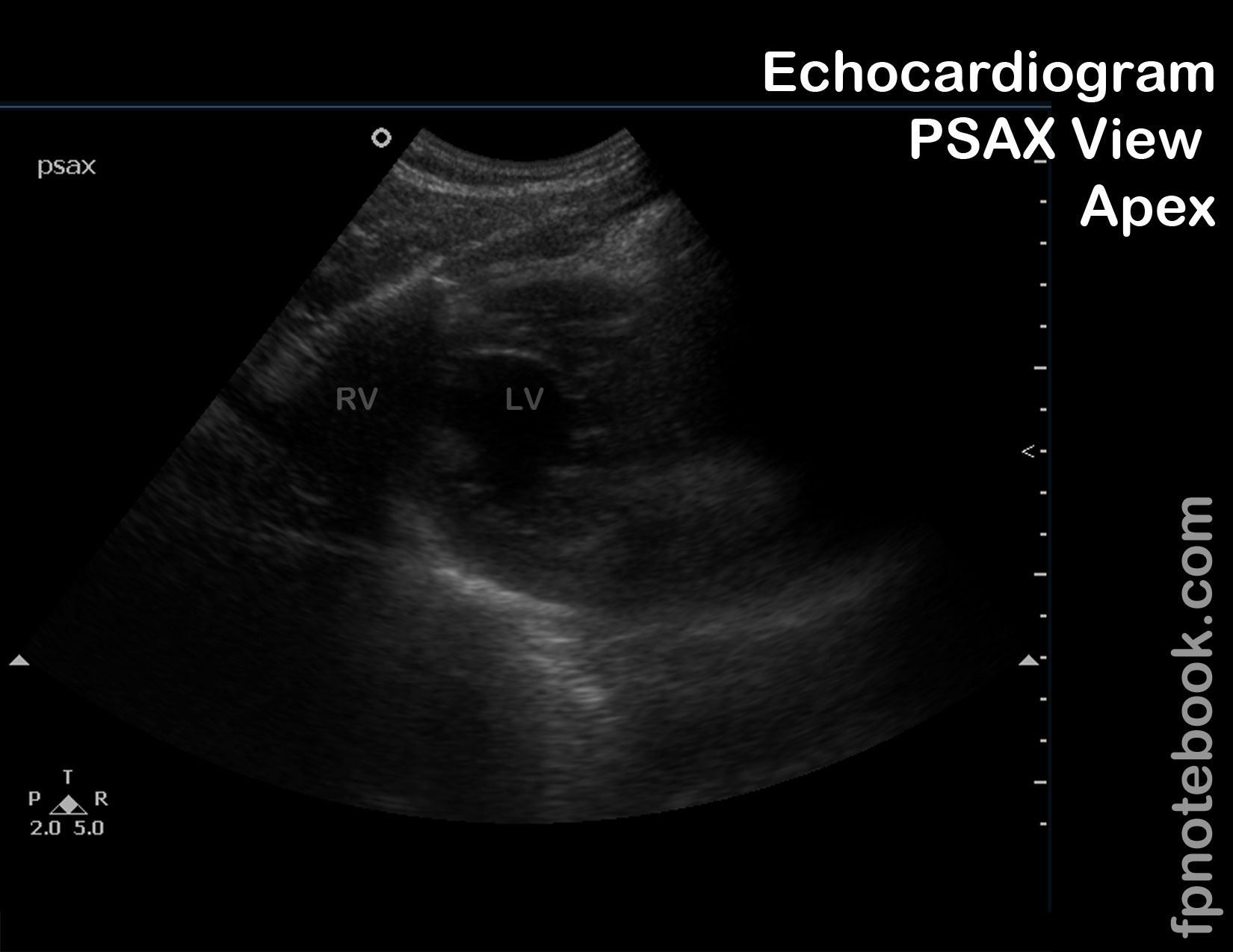
- Landmarks: Apical level
- Right ventricle (much smaller in size than left ventricle unless right ventricle dilated)
- Left ventricle (apical wall motion evaluation)
IV. Interpretation
- Bicuspid aortic valve (Aortic valve level)
- Bicuspid valve is visualized with valve open (since bicuspid valve has a fused valve leaflet that is not evident in closed position)
- Tri-leaflet appearance (Mercedes symbol) will be seen in both bicuspid and tricuspid valve when valve closed
- Left ventricle wall motion abnormality (mid-ventricle level)
- Best view to see all left ventricle walls
V. Resources
- Parasternal Short Axis View Video (SonoSite)
- Echocardiographer
VI. References
- Palma, Bourque and Jordan (2019) Introduction to Adult Echo Ultrasound Conference, GulfCoast Ultrasound, St. Petersburg
- Mateer and Jorgensen (2012) Introduction and Advanced Emergency Medicine Ultrasound Conference, GulfCoast Ultrasound, St. Pete's Beach
- Noble (2011) Emergency and Critical CareUltrasound, Cambridge University Press, New York, p. 61-88
- Orman, Dawson and Mallin in Majoewsky (2013) EM:Rap 13(1): 4-6
- Reardon (2011) Pocket Atlas Emergency Ultrasound, McGraw Hill, New York, p. 61-106