
II. Technique
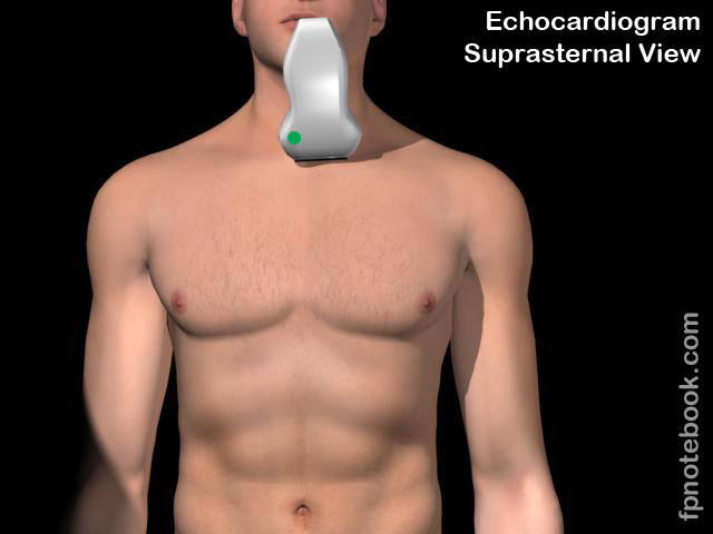
- Transducer orientation
- Transducer placed in suprasternal notch
- Transducer indicator pointed towards 9:00
- Align energy inferiorly (towards feet) until arch comes into view
- Rotate transducer clockwise until arch is in full view (12:00 to 2:00)
- May need to rotate transducer counter-clockwise
- Landmarks
- Brachiocephalic artery (right), Left Carotid Artery, Left subclavian artery
- Aortic arch
- Right pulmonary artery
- Left atrium
- Images

III. Interpretation
- Normal aorta diameters (adults)
- Proximal aorta: 4 cm
- Aortic arch: 3.5 cm
- Lower thoracic aorta (and abdominal aorta): 3 cm
- Aortic Dissection
- Aortic aneurysm
IV. Resources
- Suprasternal Notch View Video (Sonosite)
- Echocardiographer
V. References
- Mateer and Jorgensen (2012) Introduction and Advanced Emergency Medicine Ultrasound Conference, GulfCoast Ultrasound, St. Pete's Beach
- Noble (2011) Emergency and Critical CareUltrasound, Cambridge University Press, New York, p. 61-88
- Orman, Dawson and Mallin in Majoewsky (2013) EM:Rap 13(1): 4-6
- Reardon (2011) Pocket Atlas Emergency Ultrasound, McGraw Hill, New York, p. 61-106