
II. Mechanism
- Direct blow or axial load injury
III. Precautions
IV. Signs
- Volar angulation of Fracture site
- Rotational deformity if oblique Fracture
- Local swelling, Bruising and tenderness overlying Fracture
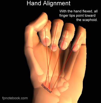
- Evaluate for malrotation (overlap deformity of affected finger when flexing fingers into a fist)
- Axes of all flexed fingers should point toward Scaphoid Bone or radial styloid (thenar eminence)

V. Imaging: XRay of Digit (AP, Lateral, Oblique)
- Evaluate for intraarticular, oblique, spliral or rotational Fractures (require orthopedic referral)
- Perform before and after manual reduction
VI. Management
- See Phalanx Fracture
- Reduction of transverse Fracture
- Apply traction away from tubercle of Scaphoid
- Flexion applied to distal fragment
- Immobilization for 4 weeks
- Splint in position of moderate flexion with ulnar gutter or radial gutter
- Open Reduction and Internal Fixation (ORIF) Indications
- Open Fracture
- Unstable Fractures (e.g. oblique, spiral, comminuted, rotational or intraarticular)
- Transverse Fracture with >2 mm displacement
- Angulation or malrotation >10 degrees
VII. References
- Perkins (2020) Crit Dec Emerg Med 34(10): 10-1