
II. Mechanism
- Direct blow or axial load injury
III. Signs
- Volar or dorsal angulation
- Local swelling, Bruising and tenderness overlying Fracture
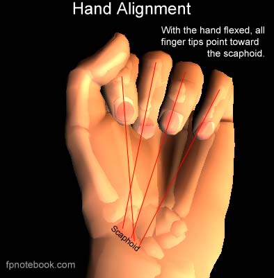
- Evaluate for malrotation (overlap deformity of affected finger when flexing fingers into a fist)
- Axes of all flexed fingers should point toward Scaphoid Bone or radial styloid (thenar eminence)

IV. Imaging: XRay of Digit (AP, Lateral, Oblique)
- Evaluate for intraarticular, oblique, spliral or rotational Fractures (require orthopedic referral)
- Perform before and after manual reduction
V. Management: Minimally Angulated, Extraarticular Fractures NOT Requiring Reduction
- Indications
- Minimal angulation (<10 degrees) AND
- Minimal to no displacement AND
- Extraarticular Fracture
- Management
- Buddy taping (between IP joints) to the adjacent finger for 3 to 4 weeks
- Aluminum splint and refer if any concerns for more complicated Fractures (see below)
- Repeat evaluation at 7 to 10 days to confirm alignment, then again at 3-4 weeks
VI. Management: Fractures Requiring Reduction
- Reduction
- Anesthesia: Digital Block or Hematoma Block
- Reduce by traction and manipulation of finger
- Immobilization after successful reduction
- Dorsal aluminum splint in extension for 6 weeks, then buddy taping for an additional 6 weeks OR
- Consider initial radial gutter or Ulnar Gutter Splint in complicated or unstable Fractures
- Post-Reduction Assessment
- Follow-up
- Repeat XRay at 7 to 10 days to confirm alignment
- Follow-up every 2 weeks
- Anticipate at least 4 to 6 weeks for healing
- Orthopedic referral indications