II. Anatomy: Anterior Abdominal Wall Muscles and Aponeuroses
III. Anatomy: Anterior Abdominal Wall Nerves
- Origin
- T6 to L1 Spinal Nerve Ventral Rami
- Upper Abdominal Wall Innervation (T6 to T12)
- Anterior Cutaneous Branches
- Lateral Cutaneous Branches
- Lower Abdominal Wall Innervation (L1)
- L1 nerve bifurcates into iliohypogastric nerve and ilioinguinal nerve
- Iliohypogastric nerve passes through external oblique superior to Superficial Inguinal Ring
- Ilioinguinal nerve passes through Inguinal Canal and out the Superficial Inguinal Ring
IV. Anatomy: Images
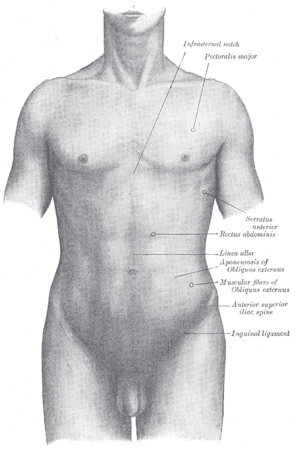
- Trunk
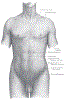 Lewis (1918) Gray's Anatomy 20th ed (in public domain at Yahoo or BartleBy)
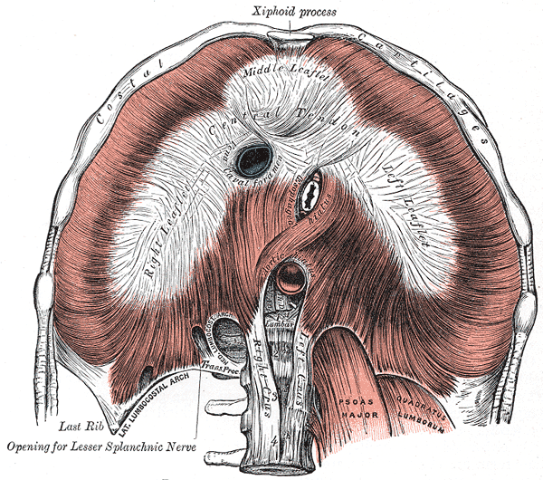
- Diaphragm
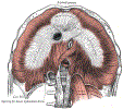 Lewis (1918) Gray's Anatomy 20th ed (in public domain at Yahoo or BartleBy)
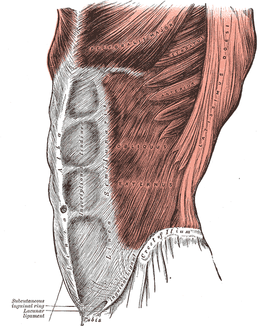
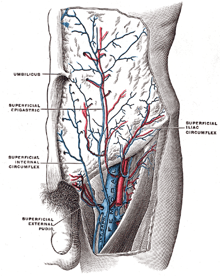
- Abdominal Wall
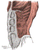 Lewis (1918) Gray's Anatomy 20th ed (in public domain at Yahoo or BartleBy)
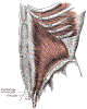 Lewis (1918) Gray's Anatomy 20th ed (in public domain at Yahoo or BartleBy)
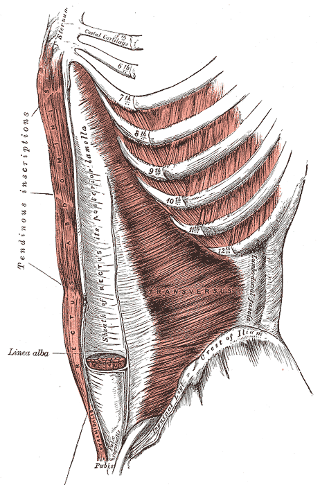
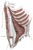 Lewis (1918) Gray's Anatomy 20th ed (in public domain at Yahoo or BartleBy)
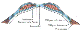 Lewis (1918) Gray's Anatomy 20th ed (in public domain at Yahoo or BartleBy)
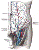 Lewis (1918) Gray's Anatomy 20th ed (in public domain at Yahoo or BartleBy)
-
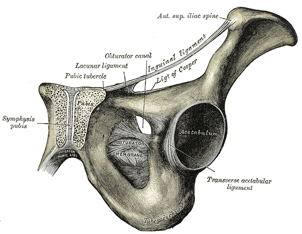
Pelvis
-
 Lewis (1918) Gray's Anatomy 20th ed (in public domain at Yahoo or BartleBy)
-