
II. Definitions
- T-Lymphocytes (T-Cells)
- T-Cells are responsible Cell-Mediated Immunity
- T-Cells are named for where they mature and differentiate (Thymus)
- T-Cells target Intracellular Pathogens (e.g. viruses and Intracellular Bacteria) and cancer cells
- Humoral Immunity (antibodies) are in contrast unable to affect Intracellular Pathogens
III. Physiology
- T-Cells
- Derived in Bone Marrow
- Migrate to Thymus
- Maturation and Differentiation into two cell lines with different T-Cell Receptors (CD4 and CD8)
- Release into peripheral circulation
- T-Cell Surface Receptors and Accessory Molecules
- T-Cell Receptors (TCR)
- Bind the Antigen on the Antigen Presenting Cell (APC)
- Similar structure to Antibody
- Each chain (a, b, g or d) has a constant region (C) and a variable region (V)
- Variable region (V) on each chain forms Antigen binding site
- TCR Types (based on polypeptides ab or gd)
- TCR-alpha-beta (TCRab+)
- TCR gamma-delta (TCRgd+)
- Plasticity
- T-Cell Receptors (TCR) have variable regions whose genes may be rearranged
- Rearranging variable region genes allows for a reflex change in TCR binding target
- Similar mechanism to B-Cell Immunoglobulin Gene modification
- T-Cell Signal Transduction related Costimulatory Receptors (Allows for T-Cell Activation)
- CD3 and Zeta (Z) are part of T-Cell Receptor Complex (along with TCR)
- CD3 and Zeta (Z) are involved with signal transduction from T-Cell surface to inside of T-Cell
- T-Cell Receptors together with CD3 and Zeta (Z) are known as T-Cell Receptor Complex
- T-Cell Receptor Complex binds Peptide-MHC Complex as first signal in T-Cell Activation
- CD28
- Binds Ligand B7 (receptor found on APCs) as part of second signal in T-Cell Activation
- CD3 and Zeta (Z) are part of T-Cell Receptor Complex (along with TCR)
- T-Cell Co-Receptors
- CD4 binds peptide/Antigen-MHC Class 2 complex on surface of Antigen Presenting Cells (APC)
- CD8 binds peptide/Antigen-MHC Class 1 complex on surface of Antigen Presenting Cells (APC)
- Adhesion Molecules (T Cell Surface Receptor Proteins that bind Ligands on Antigen Presenting Cell)
- T-Cell Receptors (TCR)
- Naive T-Cell Activation
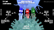
- T-Cell Receptor (TCR) binds to MHC-Antigen complex on Antigen Presenting Cells (APC)
- T-Cell Surface CD28 binds to B7 Ligand on Antigen Presenting Cell
- T-Cell Surface LFA-1 (Lymphocyte Function Associated Antigen) binds ICAM1 on Antigen Presenting Cells
- Interleukin-2 (IL2) produced by naive T Cells
- Stimulate T Cell proliferation

IV. Types: T-Cells
- Effector Cells
- T-Helper Cells (CD4+ Cells)
- T-Helper Cell Proliferation and Activation is stimulated by Antigen Presenting Cells (see above)
- Binding to T-Cell Surface Receptors and Accessory Molecules AND
- Interleukin 1 or IL-1 (released from Antigen Presenting Cells)
- T-Helper Cells then develop Interleukin 2 (IL-2) receptors once activated
- Releases Interferon
- Stimulates Phagocytosis by Macrophages
- Activates Natural Killer Cells
- Suppresses viral replication
- Releases interleuken 2
- Promotes T-Cell proliferation (including Cytotoxic T Cells, and subsequently memory cells)
- Promotes B-Cell proliferation (memory cells and plasma cells) to generate antibodies
- Additional functions
- Aid Macrophages in destroying phagocytized Bacteria
- Contribute to Graft Rejection, responding to the graft's foreign MHC, activating T-Cytotoxic Cells
- T-Helper Cell Proliferation and Activation is stimulated by Antigen Presenting Cells (see above)
- T-Cytotoxic Cells (CD8+ Cells)
- Responds to cell surface MHC I combined with degraded peptide Antigens (foreign material marker)
- Specific cytotoxic T cells form for each Antigen (e.g. virus)
- Cytotoxic T-Cell Specificity is similar to Antibody and T-Helper Cell Specificity
- Cytotoxic T Cells Target and destroy cells marked as foreign
- Tumor cells
- Virus-infected cells
- Transplanted or grafted cells (Transplant Rejection)
- Responds to cell surface MHC I combined with degraded peptide Antigens (foreign material marker)
- Memory Cells
- Cytotoxic T Cells proliferate and form Memory Cells, promoted by Helper T Cells
- T-Helper Cells (CD4+ Cells)
- Suppressor Cells
- T Suppressor Cells (Regulatory T Cells)
- Suppress immune cell activation
- Prevents autoimmune reactions by promoting self tolerance
- T Suppressor Cells (Regulatory T Cells)
- Other Cells
- Apoptosis of some cells not otherwise differentiated
V. References
- Guyton and Hall (2006) Medical Physiology, p. 419-50
- Mahmoudi (2014) Immunology Made Ridiculously Simple, MedMaster, Miami, FL