
II. Physiology
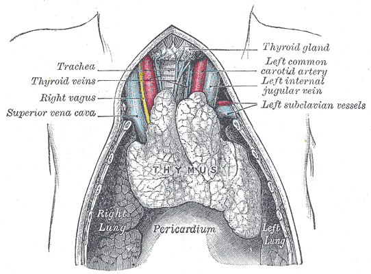
- Thymus is a bi-lobed, lymphoid gland, 3-4 cm diameter, in the anterior superior mediastinum
- Thymus primarily functions as a host for T Cell maturation and differentiation
- After forming in Bone Marrow, T Cells migrate to the Thymus for maturation
- Within the Thymus, T Cells differentiate into two cell lines with different T-Cell Receptors (CD4 and CD8)
- After maturation and differentiation, T Cells are released from the Thymus into the peripheral circulation
- T-Cells are named for their Thymus origin
- Thymus fully develops in utero, with maximal size in the first 2 years of life
- Anatomy
 Lewis (1918) Gray's Anatomy 20th ed (in public domain at Yahoo or BartleBy)
III. Imaging: Thymus Shadow on XRay
- Thymus appearance on Chest XRay
- Triangular mass in the upper chest, anterior-superior mediastinum
- Increases in size until age 12 months, then becomes much less evident by age 2 years
- Typically larger on the right side of the upper chest (AP/PA)
- Differentiate Thymus Shadow from Cardiomegaly in infants
- See Congenital Heart Disease
- See Pediatric Congestive Heart Failure
- Findings suggestive of cadiomegaly
- Boot-shaped heart (AP/PA)
- Posterior heart shadow edge overlap with Vertebral bodies (lateral)
- Anterior tracheal line displaced by posterior heart border (lateral)
IV. Differential Diagnosis: Lesions Similar to Thymus Shadow on XRay (children age >2 years, teens and adults)
- See Mediastinal Mass
- See Wide Mediastinum on Chest XRay
- Thymus Shadow is not normally seen on Chest XRay in age >2-3 years
- Mnemonic: Anterior mediastinum (retrosternal) clear space may be obscured by "Great White and the 4Ts"
- Benign Thymus disorders
- Thymic hyperplasia
- Thymic cyst
- Thymolipoma
- Thymic Lymphoid Hyperplasia (Myasthenia Gravis)
- Thyroid Goiter
- Malignancy
- Thymoma
- Thymic carcinoma
- Hodgkin Lymphoma
- Non-Hodgkin's Lymphoma
- Teratoma
- References
V. Resources
- Thymus Anatomy (StatPearls)
- Thymus (Radiopaedia)