
II. Pathophysiology
- See Bony Pelvis
-
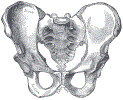
Pelvis is composed of five bony regions (ilium, ischium, pubis, Sacrum, Coccyx) held together by strong ligaments
- Significant force is required to result in Fracture of ligament disruption
- Pelvic Fractures are associated with significant bleeding
- Venous plexus that overlies the posterior arch of the Pelvis are at risk for tearing
- Fractured Pelvic Bones may also bleed significantly
- Pelvic Fracture bleeding is retroperitoneal and may be occult by external exam
- Mechanisms have a bimodal distribution
- Young Men in high energy Trauma accidents
- Elderly women with Osteoporosis, low energy mechanism (e.g. fall from standing)
- Images
 Lewis (1918) Gray's Anatomy 20th ed (in public domain at Yahoo or BartleBy)

III. Types: General
- Single Bone Pelvic Fractures
- Most common (esp. Pubic Ramus Fracture)
- See Pubic Ramus Stress Fracture
-
Acetabular Fracture
- See Acetabular Fracture
- Less, common
- Typically involve posterior acetabulum
- Pelvic Ring Fracture
- Associated with highest mortality (venous plexus and arterial related Hemorrhage)
- Divided into 3 categories (Young-Burgess Classification System) - see below
IV. Types: Unstable, Pelvic Ring Fracture Patterns (associated with other injuries)
-
General
- Young-Burgess Classification System categorizes Pelvic Ring Fractures
- Lateral Compression Pelvic Fracture
- Mechanism: Motor Vehicle Accident (e.g. rollover MVA)
- Pubic Ramus Fracture is most common manifestation
- Bladder injury or Urethral disruption are most common associated injuries
- Anterior Compression Pelvic Fracture (Open Book Fracture)
- Mechanism: Pedestrian struck by Motor Vehicle Accident
- Symphysis PubisFracture (anterior compression) with displacement is most common manifestation
- Fracture through bilateral ischiopubic rami (or Symphysis Pubis) AND SI joints
- Associated injuries
- Thoracic aorta rupture
- Sacroiliac joint opening and venous plexus disruption
- Marker for significant multisystem Trauma (due to force) such as Closed Head Injury
- Vertical Shear Pelvic Fracture
- Mechanism: High force injury (e.g. MVA or fall from height)
- Malgaigne Fracture: Ipsilateral anterior and posterior Fractures
- Anterior Fracture (ischiopubic rami or Symphysis Pubis) AND
- Posterior Fracture (SI joint, Sacrum)
- Unstable Pelvis
- Vertical displacement may be apparent on exam of the Symphysis Pubis
- Associated with significant gastrointestinal and genitourinary injuries
- Bucket Handle Fracture
- Straddle Fracture
- Bilateral ischial and pubic rami Fractures
- Results in a free floating Pubic Symphysis
V. Types: Stable Pelvic Fractures
- Stable Fractures external to pelvic ring
- Stable Fractures within the pelvic ring
- Two ipsilateral pubic or ischial ramus Fractures
- Sacroiliac joint subluxation
- Symphysis Pubis Subluxation
- Displacement >2.5 cm is unstable
VI. Precautions
- All Pelvic Fractures (and Femur Fractures) risk signficant Hemorrhage (even those that are minimally displaced)
- All Pelvic Fractures are a risk for urologic, gastrointestinal and retroperitoneal injuries
- Pelvic Fracture may give a false positive Diagnostic Peritoneal Lavage (rarely done in U.S.)
VII. Exam
- Perform a complete Trauma Examination
- See Primary Survey
- See Secondary Survey
- Compress the Pelvis by pushing both iliac crests together with force
- Assess for anterior or posterior Pelvis injury
- If the Pelvis moves inward on compression, hold this position and apply a Pelvic Binder for stabilization
- Do not repeat this exam in an unstable Pelvis (keep bound)
- Perform a careful distal CMS exam
- Distal extremity circulation (pulses, Capillary Refill)
- Distal Motor Exam
- Distal Sensory Exam
- Other examination
- Abdominal exam
- Associated lower limb Fractures
- Perineal exam for Ecchymosis
- Rectal Examination (gross blood, tone, Sensation)
- Vaginal and pelvic exam in all women with Pelvic Fracture
- Male Genitourinary Trauma (blood at Urethral meatus, perineal Ecchymosis, boggy Prostate)
- Perform Retrograde Urethrogram to exclude Urethral Trauma if external findings
- Perfrom cystogram if urethrogram negative
VIII. Imaging
-
FAST Exam (for Hemorrhage)
- Indicated in all Unstable Patients with suspected Pelvic Fractures
- High False Negative Rate for Hemoperitoneum (e.g. may miss retroperitoneal Hematoma)
-
CT Abdomen and Pelvis
- Defines Pelvic Fracture
- Defines associated genitourinary and intestinal injuries
- Other imaging and diagnostic modalities
- Pelvis XRay
- Identifies 90% of bony pelvic injuries
- Poorly predicts bleeding extent (based on Fracture appearance or type)
- Obtain if performing other bedside XRays if there is a delay for CT Pelvis
- May be sufficient in stable Trauma patients with benign Abdomen and Pelvis
- Minimum imaging in Unstable Patients with positive FAST requiring emergent Trauma surgery
- Diagnostic Peritoneal Lavage
- Completely replaced by CT Abdomen and Pelvis in the United States
- Retrograde Urethrogram (followed by cystogram if negative) Indications
- Men with blood at Urethral meatus or boggy Prostate
- Gross Hematuria
- Voiding difficulty
- Perineal Bruising
- Pelvis XRay
IX. Evaluation
- Stable: CT Abdomen and Pelvis
- Injury or peritonitis in addition to Pelvic Fracture requiring laparotomy
- Laparotomy for other indication and visualize Pelvic Fracture region at same time
- Isolated Pelvic Fracture
- Evaluation by Trauma surgery
- Intervention Radiology (angiography) for concerning findings (e.g. soft tissue blush or Hematoma near Fracture site)
- Injury or peritonitis in addition to Pelvic Fracture requiring laparotomy
- Unstable: FAST Exam
- FAST Positive for Hemorrhage
- Emergent Laparotomy to identify and manage bleeding source
- Source may be from concurrent Liver Laceration, Ruptured Spleen or Mesenteric Artery bleeding
- FAST Negative for Hemorrhage
- Blood Transfusion (see below)
- Consider Intervention Radiology to identify bleeding source (and consider internal iliac embolization)
- Consider Laparotomy for persistent instability if above measures are unsuccessful
- Consider Resuscitative Endovascular Balloon Occlusion of the Aorta (REBOA)
- See management below
- FAST Positive for Hemorrhage
X. Management
-
Pelvic Binder (e.g. T-POD or bed sheet)
- See Pelvic Binder
- Provides pain relief and Fracture stabilization (similar to external fixation)
- Does not reduce Hemorrhage significantly (although may reduce Blood Transfusion requirements)
- Does not affect arterial bleeding
- Manage Hemorrhagic Shock
- Start replacing Blood Products early
- Replace Red Blood Cells (as well as Platelets and Fresh Frozen Plasma 1 unit/unit pRBC)
- Emergent surgical Consultation
- Trauma surgery, orthopedics, general surgery or urology depending on extent of injuries
- Determine management (laparotomy, Intervention Radiology or observation)
- Surgical management options (both followed by angiography by Intervention Radiology)
- Laparotomy with direct packing and possibly internal iliac artery ligation OR
- Preperitoneal packing via short suprapubic space incision
- Indicated only if other Hemorrhage sources have been excluded
- Trauma surgery, orthopedics, general surgery or urology depending on extent of injuries
- Angiography by Intervention Radiology indications
- See Evaluation above
- Indicated for persistent bleeding from Pelvic Fracture
- Do not delay emergent surgery in an Unstable Patient if angiography is not immediately available (e.g. <30 min)
- Managed with pelvic embolization (typically internal iliac embolization)
- Other possible measures
- Resuscitative Endovascular Balloon Occlusion of the Aorta (REBOA)
- Percutaneous balloon delivered via groin catheter and inflated in aorta above level of Hemorrhage
- Indicated in severe Pelvic Fractures without Cardiac Arrest who need immediate temporizing measures
- May be considered in Peri-Arrest patient without obvious source of Hemorrhage
- Best used for short-term bridging to definitive procedure (risk of distal ischemia)
- Resuscitative Endovascular Balloon Occlusion of the Aorta (REBOA)
- Ineffective Measures
- Bladder Distention with foley (Bladder is too anterior to provide adequate posterior compression)
- Stable Fractures not requiring surgery
- Pubic Ramus Fracture
- Anterior-Posterior Compression (APC) Fracture Type 1
- Lateral Compression Fracture Type 1
XI. Prognosis: Acute Mortality
- Mortality 15-40% for an isolated Pelvic Fracture with secondary bleeding and Hypotension
- Mortality 50% for a Pelvic Fracture AND intraabdominal injury
- Mortality 90% for a Pelvic Fracture AND intraabdominal injury AND Head Injury
XII. References
- Eiff (1998) Fracture Management for Primary Care, p. 174-7
- Inaba in Herbert (2013) EM:Rap 13(11): 3-4
- Inaba and Herbert in Herbert (2014) EM:Rap 14(4): 10-11
- Orman and Hicks in Herbert (2017) EM:Rap 17(2): 8-9
- Perkins (2022) Crit Dec Emerg Med 36(6): 18-9 [PubMed]