
II. Definitions
- Wolff-Parkinson-White Syndrome
- Arrhythmia associated with AV bypass tract (accessory path outside the AV nodal path)
- Subtype of Atrioventricular Reciprocating Tachycardia (AVRT) distinguised by its delta wave
III. Epidemiology
- Prevalence: 2 per 1000 general population
IV. Pathophysiology
- Subtype of Atrioventricular Reciprocating Tachycardia (AVRT)
- Delta wave and Short PR Interval
- Prolonged QRS Duration
- Anterograde conduction via the accessory path
- Atrioventricular bypass tract
- Circumvents normal PR Interval delay (up to 0.2 sec)
- Allows for ventricular pre-excitation
- Predisposes to 3 classic Dysrhythmias
- Orthodromic Atrioventricular Reciprocating Tachycardia (AVRT, 65-80%, narrow complex)
- See Orthodromic AVRT
- WPW-Related Paroxysmal Atrial Fibrillation (20-25%)
- Wide Complex Tachycardia often with atypical appearing QRS Complexes
- QRS Complexes show beat-to-beat variability in morphology, amplitude and width
- Narrow QRS Complexes may be intermittently seen
- QRS results from the fusion of the accessory and AV nodal pathway transmissions
- Rapid ventricular rates often >220 bpm
- Irregularly irregular rhythm
- Wide Complex Tachycardia often with atypical appearing QRS Complexes
- Antidromic Atrioventricular Reciprocating Tachycardia (AVRT, <10%, wide complex)
- See Antidromic AVRT
- Orthodromic Atrioventricular Reciprocating Tachycardia (AVRT, 65-80%, narrow complex)
V. Findings: EKG changes
- Precautions
- WPW EKG Findings may be variably present
- Classic findings are more prominent with Valsalva Maneuver (or other increased vagal tone)
- Narrow or Short PR Interval (PR <0.12)
- Bypass tract results in faster conduction through the AV Node and earlier ventricular depolarization
- Look closely for Delta wave when a narrow PR Interval is identified on EKG
- Delta wave
- Slurred upstroke of QRS (hockey stick appearance)
- Bypass tract impulse reaches ventricular Myocardium before AV Node conduction (ventricular pre-excitation)
- Resulting ventricular impulses are cell-to-cell and slower than bundle branch conduction
- Slurred QRS appearance results from the fusion of 2 depolarization waves
- Early bypass tract impulses depolarizing ventricular Myocardium cell-to-cell (wide)
- Normal AV Node impulses depolarizing the bundle branches (narrow)
- Concealed accessory paths conduct only retrograde, and do NOT have a delta wave
- Slightly Wide QRS
- Wide QRS related to delta wave (to extent that PR Interval is narrowed)
- Pseudoischemic Changes
- Q Waves associated with abnormal depolarization
- ST Segment deviation and T Wave Inversion associated with abnormal repolarization
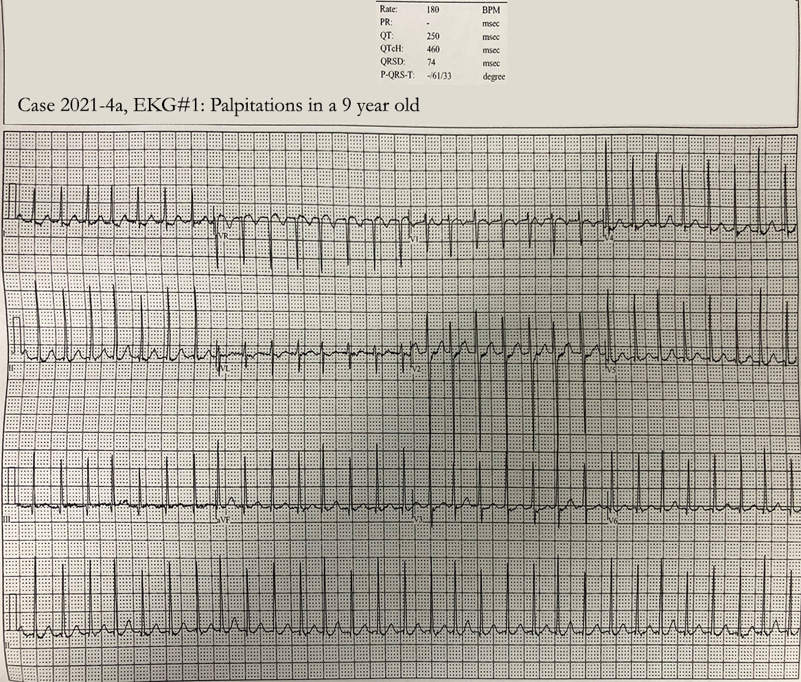
- Images
VI. Differential Diagnosis
- Right or Left Bundle Branch Block (wide complex)
- Myocardial Infarction (Q Wave when QRS negative)
VII. Precautions
- Agents to avoid in WPW (may accelerate Arrhythmia via accessory path)
- Adenosine
- Beta Blockers (e.g. Metoprolol)
- Calcium Channel Blockers (e.g. Verapamil, Diltiazem)
- Digoxin (Lanoxin)
- Have a high index of suspicion in young patients with Syncope
- WPW may be present despite an absence of Short PR Interval and a Delta Wave
-
Sinus Tachycardia can still occur with all of the typical reasons seen in patients without WPW
- Consider Dehydration, infection, Pulmonary Embolism in the differential in a patient with WPW and Tachycardia
VIII. Management
- See Unstable Tachycardia
- Safe interventions in WPW
- Synchronized Cardioversion (preferred)
- Procainamide
- Avoid AV Nodal blocking agents (esp. in WPW-Related Paroxysmal Atrial Fibrillation)
- Contraindicated AV nodal blockers include Beta Blockers, Calcium Channel Blockers, Adenosine, Amiodarone
- AV nodal blockade may potentiate the unregulated accessory pathway increasing the ventricular rate (V fib risk)
- Any negative inotrope may also worsen hemodynamic collapse
IX. Complications
- Atrioventricular Re-Entry Tachycardia (AVRT)
- Rates are typically very high (200-300 bpm)
- Reentrant Paroxysmal Supraventricular Tachycardia
- Orthodromic AVRT in most cases (Antidromic AVRT is much less common)
-
Atrial Fibrillation (20% of WPW patients)
- When associated with preexcitation, may degenerate into Ventricular Fibrillation
- Atrial Flutter (7% of WPW patients)
- Ventricular Tachycardia or Ventricular Fibrillation
- Sudden Cardiac Death
X. References
- Braude, Swadron and Orman et. al. in Herbert (2012) EM:RAP 12(7): 1-2
- Goldberger (1999) Clinical Electrocardiography, p 127-8
- Grauer (2001) 12 Lead EKG, p. 27
- Joshi and Dermark (2016) Crit Dec Emerg Med 30(8):3-12
- Layng, Vandersteenhoven, Brady (2025) Crit Dec Emerg Med 39(8): 16-7
- Vandersteenhoven, Brady (2025) Crit Dec Emerg Med 39(10): 15-7