
II. Epidemiology
- Middle aged and older adults (esp. 40 to 60 years old)
- More common in women
III. Predisposing factors
- Overuse injury in athletes
- Low cut shoes
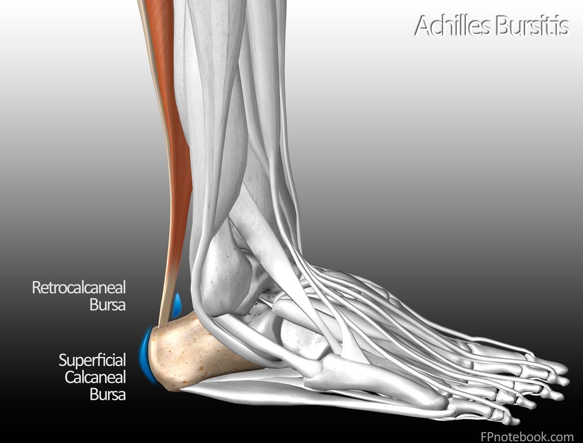
IV. Anatomy
-
General
- Two bursae located near achilles tendon insertion
- Superficial Calcaneal Bursa (Pump-Bump)
- Located over achilles tendon
- Irritated by constant rubbing by shoe
- Associated with thin heel pad
- Retrocalcaneal bursa
- Located under achilles tendon
- Irritated by Calcaneus
- Prominent posterosuperior angle (Haglund's Disease)
V. Symptoms
- Heel Pain with swelling and erythema
- Pain exacerbating factors
- Worse at the beginning of activity (e.g. walking)
- Wearing shoes may worsen pain
- Painful limp may develop
VI. Signs
- Inflammation at achilles tendon insertion on Calcaneus
- Two finger Squeeze Test
- Patient with plantar flexed ankle
- Examiner compresses tissue that is immediately anterior to the distal achilles tendon
- Positive if compression causes pain
- Images
VII. Differential Diagnosis
- See Heel Pain
VIII. Imaging
- Indicated only in refractory cases
-
XRay: Haglund's Deformity
- Bone spur on superior Calcaneus
- Calcified distal achilles tendon
-
Ultrasound
- Hyperemia in bursa region (increased Blood Flow on color doppler)
- Hypoechogenic fluid in retrocalcaneal bursa
IX. Management
-
General Measures
- Heel pads
- NSAIDs
- Alternate Ice Therapy with heat therapy
- Consider wearing sandals (open back)
- Elevation of shoe heel with soft cushion
-
Corticosteroid Injection are not recommended
- Offer only short-term pain relief (ineffective in 14% of patients)
- Exercise caution with local steroid injections (and use Ultrasound guidance if performed)
- Risk of Achilles Tendon Rupture or weakening in the first 6 months after injection
- Surgery for refractory cases
- Bursa and bony prominence resection
- Symptom and function improvement by 4 months is typical