
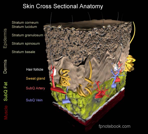
II. Anatomy
- Complete Cross Sectional Anatomy
 Lewis (1918) Gray's Anatomy 20th ed (in public domain at Yahoo or BartleBy)
- Epidermis
 Lewis (1918) Gray's Anatomy 20th ed (in public domain at Yahoo or BartleBy)
III. Definitions
- Epidermis (external skin surface)
- Keratinised squamous epithelium
- Thickness
- Eyelids: 0.05 mm
- Palms and soles: 1.5 mm
- Dermis (supports Epidermis)
- Thick, dense, fibroelastic connective tissue
- Highly vascularized
- Contains Sensory Receptors
- Hypodermis (Subcutaneous layer)
- Loose connective tissue with adipose tissue
IV. Physiology: Skin Functions
- Sensation (largest sensory organ in the body)
- Protection
- Prevents Dehydration
- Prevents infection
- Physical barrier to injury
- Protects against ultraviolet light injury (Melanin)
-
Thermoregulation
- Insulation (hair and adipose tissue)
- Heat dissipation
- Sweat evaporation
- Increased Blood Flow
- Metabolic
- Energy storage of Triglycerides in adipose tissue
- Vitamin D synthesis
V. Anatomy: Epidermis Cell Layers (cells mature from inner to outer)
- Stratum Corneum (Cornified Layer)
- Outermost layer of Epidermis
- Composed mostly of keratin (fibrous Protein)
- Cells desquamated (27 days after production)
- Stratum Lucidum (present only in very thick skin)
- Stratum Granulosum (Granular Layer)
- Darker layer with intracellular granules
- Produces keratin
- Stratum Spinosum (Prickle Cell Layer)
- Composed of Keratinocytes
- Cells produced by basal layer and growing
- Keratin production starts
- Stratum Germinativum (Stratum Basale, Basal Cell Layer)
- Innermost layer of Epidermis
- Cells are produced here in the germinal layer
- Forms the prickle cells in the layer above
VI. Anatomy: Interspersed cells and units
- See Hair Follicle
- See Sweat Gland
- See Sebaceous Gland
- See Melanocyte
- Merkel's Cell
-
Langerhans Cells (in Prickle Layer)
- Dendritic histiocytic cells
- Intercept Antigenic signal and pass to lymphoid cells
- Desmosome (Macula adherens)
- Intercellular bridge that attaches epidermal cells
- Small dense Plaque with protruding tonofilaments
VII. References
- Habif (1996) Clinical Dermatology, Mosby, p. 24
- Murphy in Cotran (1989) Robbins Pathology, p. 1277-8
- Wheater (1987) Functional Histology, p. 130-2