II. Findings: Normal Colposcopy
- Original Squamous epithelium (OSE)
- Distal Margin: Vulva
- Proximal Margin: Original Squamocolumnar junction
- Columnar epithelium (CE)
- Distal Margin: Squamous metaplasia
- Proximal Margin: Ectocervix and into Uterus
- Squamous metaplasia (SM)
- Mature Squamous Metaplasia (MSM)
- Distal Margin: Original Squamocolumnar Junction
- Proximal Margin: New Squamocolumnar junction
- Immature Squamous Metaplasia (ISM)
- Distal Margin: New Squamocolumnar junction
- Proximal Margin: Limit of squamous metaplasia
- Mature Squamous Metaplasia (MSM)
- Squamocolumnar junction (SCJ)
- Transformation zone
- Gland opening
- Nabothian cyst
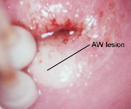
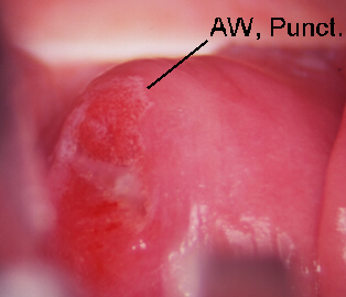
III. Findings: Changes associated with Low Grade SIL
- Koilocytosis
- Hyperkeratosis or Leukoplakia
- Light Acetowhite staining of Epithelium
- Blood vessel changes (Fine punctation or mosaicism)
- Atypical Lesion Margins: Irregular, angular, or jagged
- Condyloma or Papilloma: exophytic, verrucous, or flat
- Iodine Staining


IV. Findings: Changes Associated with High Grade SIL
- Blood Vessel changes (Coarse punctation or mosaicism)
- Abnormal blood vessels
- Erosions
- Dense Acetowhite staining of epithelium