
II. Approach: Suprapublic View (Pelvis)
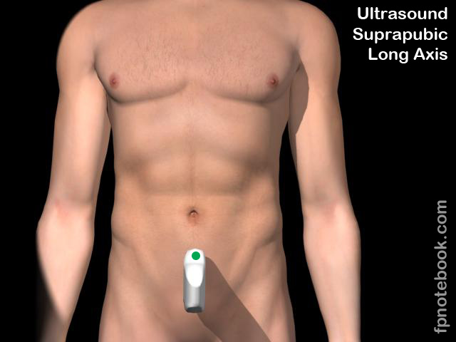
- Transducer positioning
- Placement: Low suprapubic region
- Place probe immediately above suprapubic bone
- Axis: Long axis (longitudinal)
- Probe indicator at 12:00
- Longitudinal axis is best for visualizing anatomic landmarks (easiest for those new to Ultrasound)
- Probe Direction
- Perpendicular to Pelvis - towards low Lumbar Spine
- Gain
- Turn down gain to visualize structures behind the Bladder
- Placement: Low suprapubic region
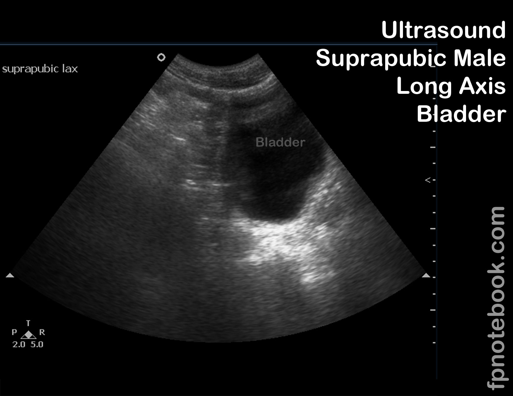
- Landmarks (based on longitudinal axis with probe indicator at 12:00)
- Bladder and Bladder midline
- Draw a line through the midline of longitudinal Bladder view from screen top to screen bottom
- Superior to line (screen left)
- Free fluid and blood collects here, deep to Bladder (this region should be key focus)
- Men collect flud immediately deep to Bladder (in rectovesical space)
- Women collect fluid deep to Uterus (deep to Bladder), in Pouch of Douglas
- Bladder Diverticulum may also form fluid pockets posterior to Bladder (False Positive FAST)
- Inferior to line (screen right)
- Represents low Pelvis structures (e.g Prostate)
- Region of little concern in the trauma Ultrasound
- Uterus
- Bladder and Bladder midline
- Conditions
- Blood in Pelvis (Douglas Pouch)
- Focus on longitudinal midline view
- Blood will appear as a dark black collection, superior to Bladder (screen left)
- Blood (or fluid) will outline omentum appearing as irregular shapes
- Contrast with normal suprapubic region appearance (gray, hazy, partially hypoechoic)
- Perform initially prior to Foley Catheter insertion (Bladder should be full to start)
- May be difficult to distinguish Bladder from blood in Pelvis
- Consider re-scanning after urine drained with Foley Catheter
- Focus on longitudinal midline view
- Younger children
- May be more sensitive than Morrison's Pouch for intraabdominal blood
- Males
- Any free fluid is abnormal
- Women
- Free fluid seen on transabdominal Ultrasound is not typically physiologic
- Contrast with Transvaginal Ultrasound which is sensitive enough to find trace fluid
- Free fluid seen on transabdominal Ultrasound is not typically physiologic
- Blood in Pelvis (Douglas Pouch)
- Images: Long Axis
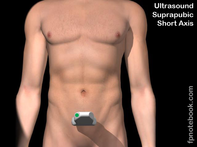
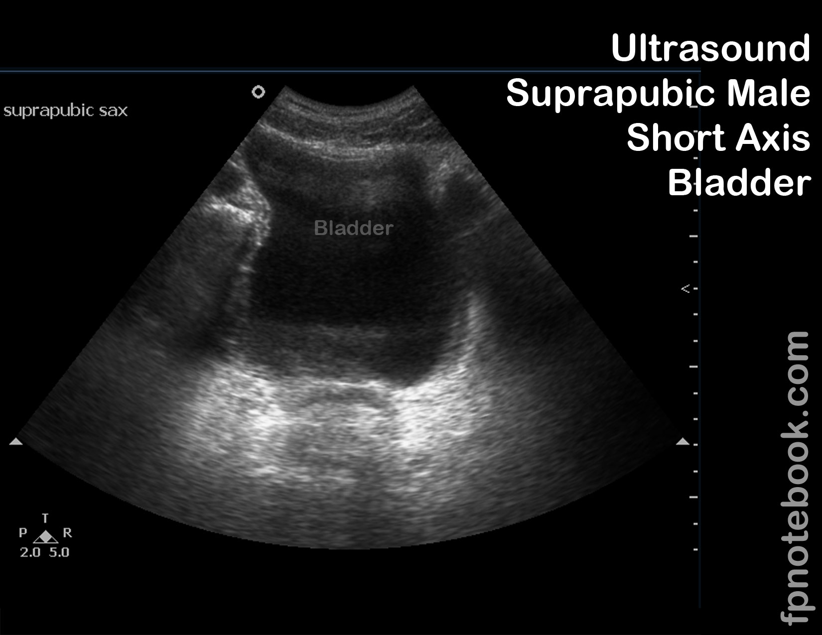
- Images: Short Axis
III. Resources
- FAST Exam Suprapubic (Dr. Mandavia,SonoSite)
- FAST Exam Female Pelvis (Dr. Mandavia, SonoSite)
- Fast Exam Male Pelvis (Dr. Mandavia, SonoSite)
IV. References
- Reardon (2016) FAST Scan, Online Video Stabroom.com, accessed 4/1/2016
- Reardon (2013) Emergency Ultrasound Course, 3rd Rock Ultrasound, Minneapolis, MN
- Alameda County Trauma Service FAST Exam
- Mateer (2012) Introduction to Trauma Ultrasound Video, GulfCoast Ultrasound, VL-95-T
- HCMC FAST Exam