
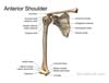
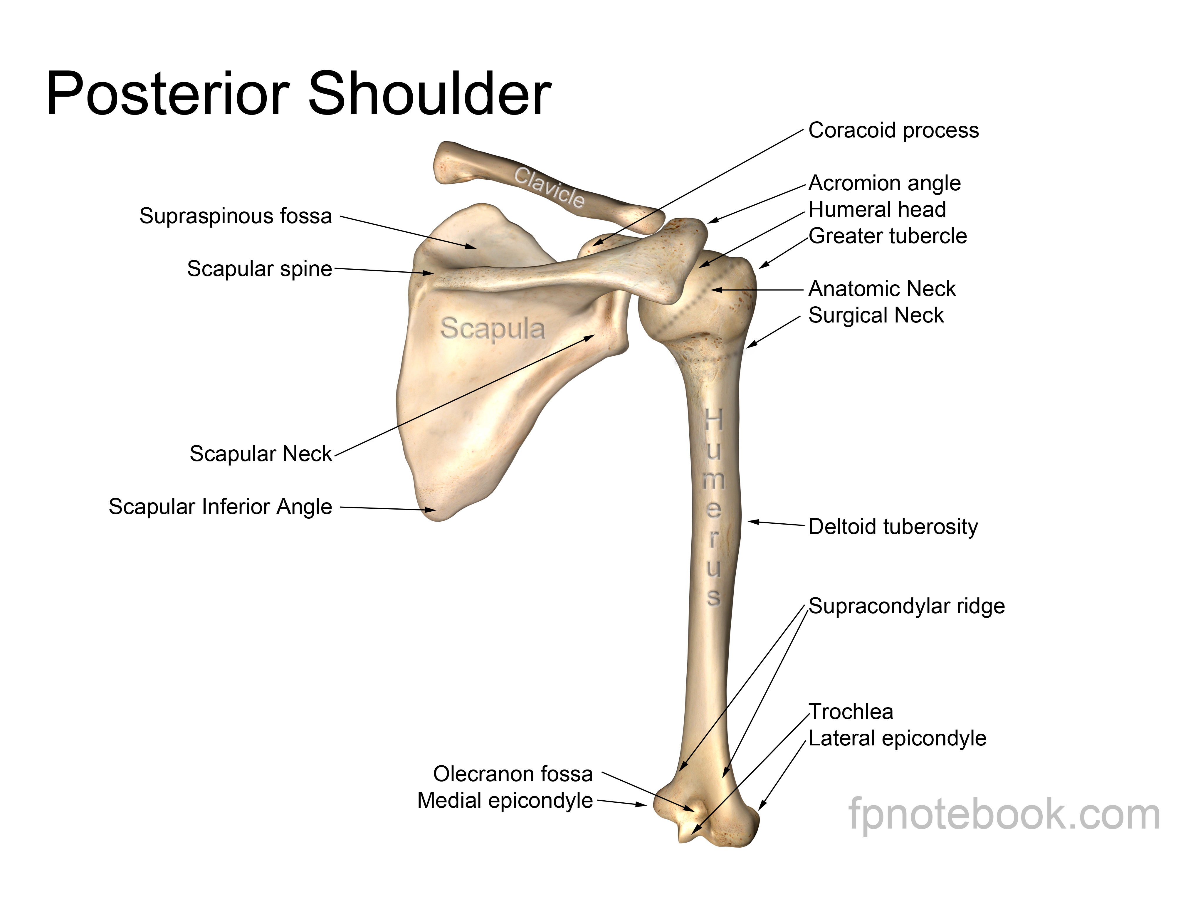
II. Anatomy
- Bone and Ligament
 Also available as a Poster size image. See printing instructions and image restrictions.
 Also available as a Poster size image. See printing instructions and image restrictions.
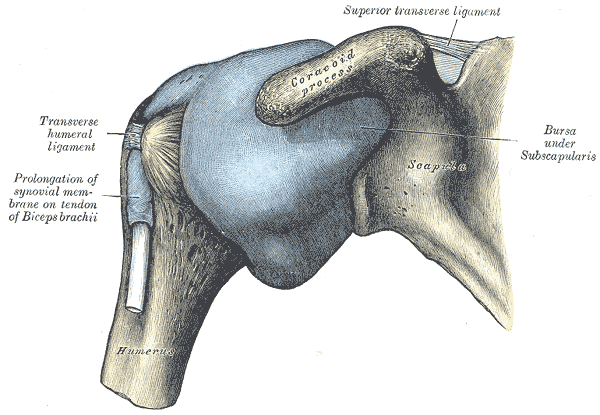
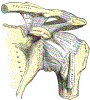 Lewis (1918) Gray's Anatomy 20th ed (in public domain at Yahoo or BartleBy)
 Lewis (1918) Gray's Anatomy 20th ed (in public domain at Yahoo or BartleBy)
 Lewis (1918) Gray's Anatomy 20th ed (in public domain at Yahoo or BartleBy)
-
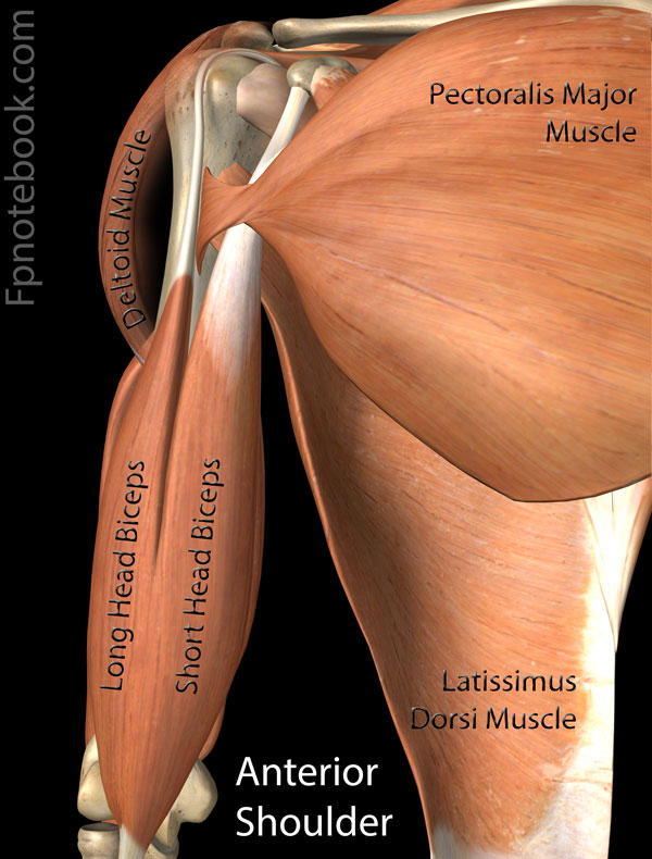
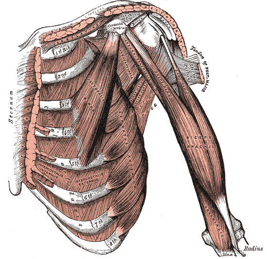
Muscles
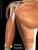
 Lewis (1918) Gray's Anatomy 20th ed (in public domain at Yahoo or BartleBy)
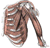 Lewis (1918) Gray's Anatomy 20th ed (in public domain at Yahoo or BartleBy)
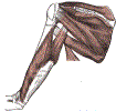 Lewis (1918) Gray's Anatomy 20th ed (in public domain at Yahoo or BartleBy)


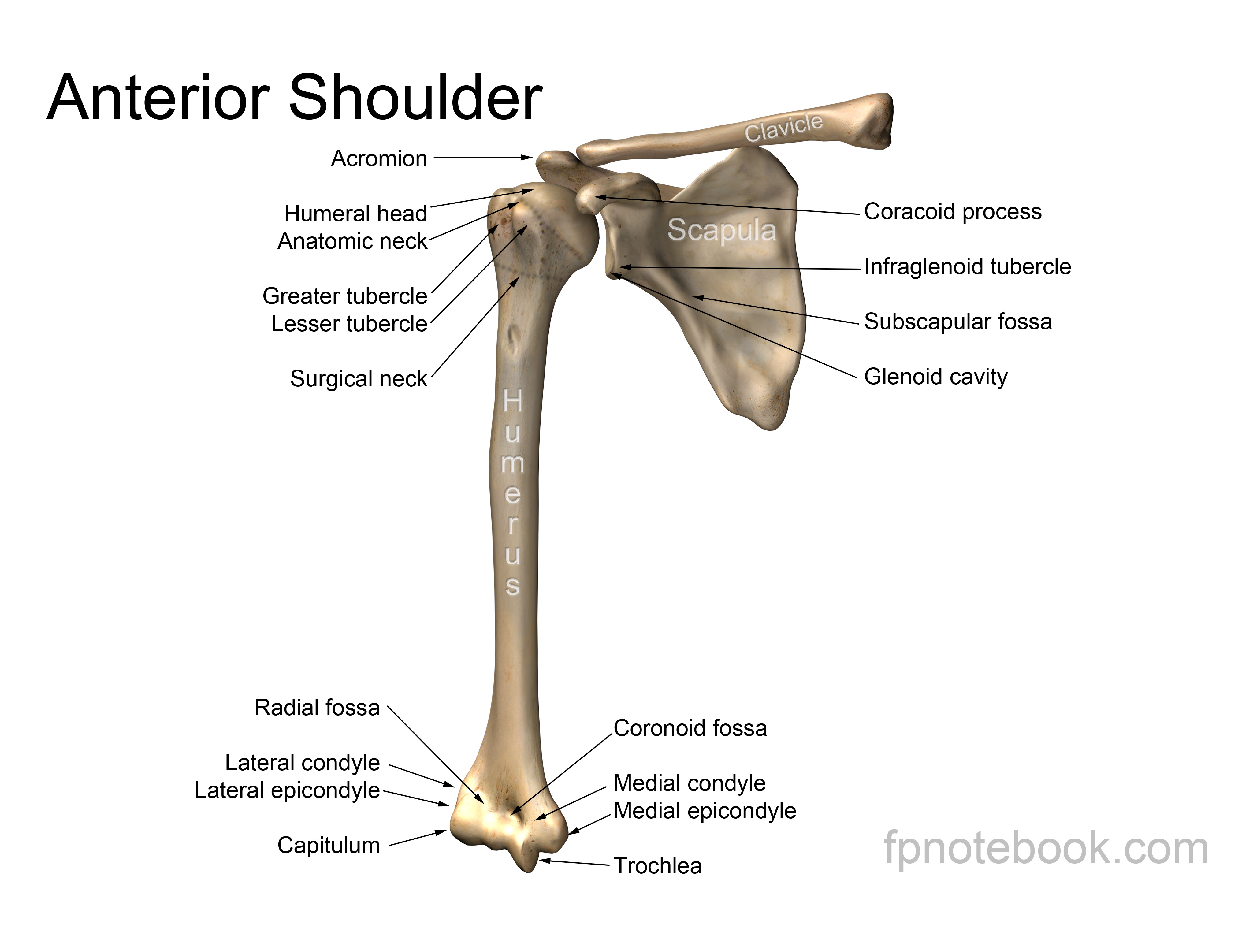
III. Anatomy: Bones
IV. Anatomy: Muscles
V. Anatomy: Joints and Articulations
- Glenohumeral joint (GH Joint)
- Glenoid of Scapula meets humeral head
- Golf ball on golf tee
- Articulation with bone: 30%
- Articulation with glenoid labrum: 75%
- Posterior Chest Wall (Scapulothoracic articulation)
- Acromioclavicular Joint (AC Joint)
- Acromion of Scapula meets the clavicle
- Sternoclavicular Joint (SC Joint)
- Four ligaments (in addition to a saddle-like structure) stabilize the joint
- Anterior sternoclavicular ligament
- Posterior sternoclavicular ligament
- Interclavicular ligament
- Costoclavicular ligament
- Four ligaments (in addition to a saddle-like structure) stabilize the joint
VI. Anatomy: Nerves
- Rotator cuff is innervated by Brachial Plexus derived nerves
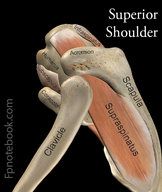
VII. Anatomy: Subacromial Space
- Coracoclavicular arch forms the roof of the subacromial space
- Coracoid process
- Coracoacromial ligament
- Acromion process
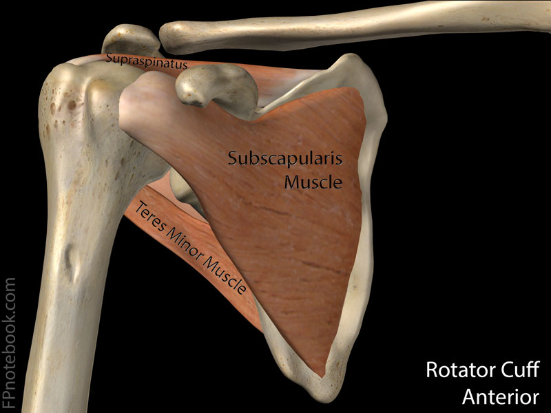
- Rotator cuff composition (mnemonic: SITS Muscles)
- Subacromial bursae
- Separates cuff from overlying coracoacromial arch
- Includes subdeltoid bursa and subcoracoid bursa
- Image
VIII. Physiology: Function
- Scapula stabilized prior to arm movement
- Rotator Cuff (Musculotendinous)
- Steadies humeral head in glenoid
- Results in humeral head descent
- Scapulothoracic Musculature
- Results in forward and lateral Scapula movement
- Initiates first 60-70 degrees of Shoulder Abduction
- Glenohumeral Musculature
- Deltoid and supraspinatus Muscles
- Accounts for last two thirds of Shoulder Abduction
IX. Physiology: Muscle action at Glenohumeral joint
- Shoulder Abduction
- Shoulder Adduction
- Shoulder Flexion
- Shoulder Extension
- Shoulder Internal Rotation
- Shoulder External Rotation
Images: Related links to external sites (from Bing)
Related Studies
| Definition (SCTSPA) | Región del cuerpo definida por la articulación del hombro y las estructuras circundantes |
| Definition (SNOMEDCT_US) | The body part defined by the shoulder joint and its surrounding structures |
| Definition (NCI) | The region of the body between the neck and the upper arm. |
| Definition (NCI_CDISC) | The region of the body between the neck and the upper arm. (NCI) |
| Definition (CSP) | junction of the arm and trunk; also that part of the trunk which is bounded at the back by the scapula. |
| Concepts | Body Location or Region (T029) |
| MSH | D012782 |
| SnomedCT | 16982005 |
| HL7 | SHOL, SHOLJ |
| LNC | LP7584-8, MTHU002689, LA4298-1 |
| English | Shoulder, Shoulders, Shoulder region, Structure of shoulder region, Structure of shoulder region, unspecified, anatomies shoulder, shoulder anatomy, shouldering, shoulder, anatomy shoulder, shoulders, SHOULDER, Shoulder region structure, Shoulder region structure (body structure), Shoulder, NOS, Shoulder (Anatomy), Sholder Joint |
| Swedish | Skuldra |
| Spanish | estructura de la región de la espalda, estructura de la región del hombro, estructura de la región del hombro (estructura corporal), estructura de región de hombro (estructura corporal), estructura de región de hombro, Hombros, hombro, estructura de la región de la espalda (estructura corporal), Hombro |
| Czech | rameno |
| Finnish | Olkapää |
| Russian | PLECHO, ПЛЕЧО |
| Croatian | RAME |
| Latvian | Plecs |
| Polish | Okolica naramienna, Bark, Obręcz kończyny górnej |
| Norwegian | Skulder |
| Portuguese | Ombros, Ombro |
| French | Épaule |
| German | Schulter |
| Italian | Spalla |
| Dutch | Schouder |
Ontology: Shoulder Injuries and Disorders (C1456701)
| Definition (MEDLINEPLUS) |
Your shoulder joint is composed of three bones: the clavicle (collarbone), the scapula (shoulder blade), and the humerus (upper arm bone). Your shoulders are the most movable joints in your body. They can also be unstable because the ball of the upper arm is larger than the shoulder socket that holds it. To remain in a stable or normal position, the shoulder must be anchored by muscles, tendons and ligaments. Because the shoulder can be unstable, it is the site of many common problems. They include sprains, strains, dislocations, separations, tendinitis, bursitis, torn rotator cuffs, frozen shoulder, fractures and arthritis. Usually shoulder problems are treated with RICE. This stands for Rest, Ice, Compression and Elevation. Other treatments include exercise, medicines to reduce pain and swelling, and surgery if other treatments don't work. NIH: National Institute of Arthritis and Musculoskeletal and Skin Diseases |
| Concepts | Injury or Poisoning (T037) |
| English | Shoulder Injuries and Disorders |
