
II. Indication
- Aspiration
- Knee Effusion of unclear etiology
- Evaluation for Septic Knee
- Injection (Synvisc or Corticosteroid)
- Knee Osteoarthritis
- Gouty Arthritis affecting the knee
- Pseudogout affecting the knee
III. Preparation
- Needle
- Gauge: 25-27 (injection), 18-20 (aspiration)
- Length: 1.5 inches
- Syringe: 10 ml (injection) or 30-60 ml (aspiration)
-
Corticosteroid options
- Betamethasone (Celestone) 1 to 2 ml of 6 mg/ml
- Methylprednisolone (Depo-Medrol): 1 to 2 ml of 40 mg/ml
- Dexamethasone 8 mg
- Triamcinolone 40 mg
-
Anesthetic: 5 to 7 ml
- Lidocaine 1% OR
- Bupivacaine 0.25% or 0.5%
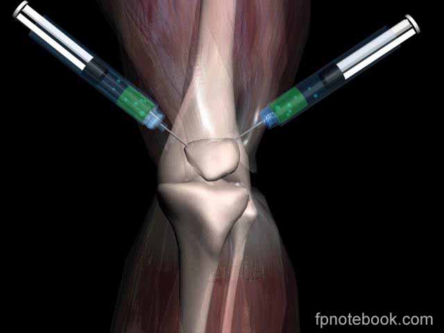
IV. Technique: Native Knee
- Images
- Patient position
- Patient supine with knee in slight flexion (15 to 20 degrees)
- Prop up knee on towel roll in popliteal space
- Sterilize local skin with Betadine or Hibiclens
-
Ultrasound guidance (optional)
- Linear probe 12 MHz
- Place probe at the lateral superior aspect of the Patella (see landmarks below)
- Direct probe medially
- Target is the suprapatellar pouch
- Mark needle insertion site based on approach
- Lateral Suprapatellar Approach
- Medial Suprapatellar Approach
- Knee flexed 60-90 degrees
- Insert needle medial to Patellar tendon
- Stay parallel to tibial plateau
- Risk of Meniscus Injury
- Medial Approach
- Risk of needle injury to the medial meniscus (uncommon)
- Aspirate first, then inject
- Inferior Approach
- Aspirate first and then inject
- Use first syringe to aspirate joint contents
- Use hemostat to detach syringe from needle
- Attach syringe with Corticosteroid
- Inject Corticosteroid mix into joint
V. Technique: Prosthetic Knee
- See Periprosthetic Joint Infection
- Indications
- Suspected Periprosthetic Joint Infection after total knee arthroplasty (TKA)
- Preparation
- Patient supine with affected knee in slight flexion
- Procedure
- Use sterile technique
- Ultrasound linear probe
- Apply Ultrasound probe sterile cover
- Start with probe indicator oriented toward patient's head
- Place the probe in the prepatellar fossa
- Slide the probe superiorly until the effusion is visible (in the suprapatellar space)
- Identify landmarks (Quadriceps femoris tendon, Femur, Fat pad)
- Rotate the probe 90 degrees to allow for in-plane needle insertion
- Prepare needle insertion site
- Chlorhexidine
- Inject Local Anesthetic at skin surface (e.g. Lidocaine 1 to 2%)
- Avoid injecting into effusion
- Insert and advance an aspiration needle toward effusion (e.g. 18 gauge)
- Use In-Plane technique to follow the needle tip into effusion
- Target effusion will be deep to the quadriceps tendon
- References
- Voorhees and Riveros (2024) Crit Dec Emerg Med 38(3):22-3
VI. References
- Pfenninger (1994) Procedures, p. 1036-54
- Cardone (2003) Am Fam Physician 67(10):2147-52 [PubMed]
- Webb (2024) Am Fam Physician 109(1): 61-70 [PubMed]
- Zuber (2002) Am Fam Physician 66(8):1497-1500 [PubMed]