
II. Definition
- Meninges
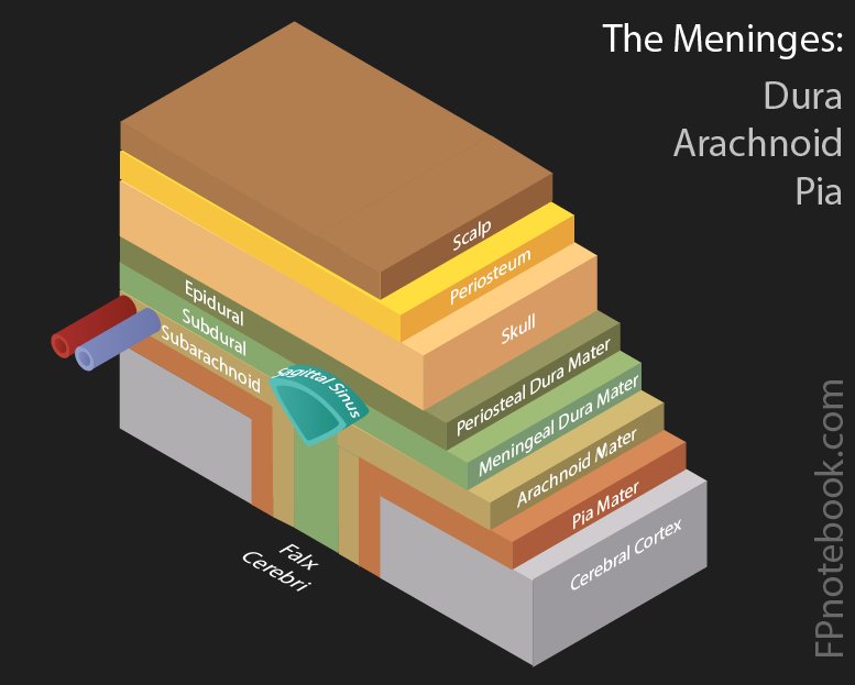
- Membranous lining (3 layers, dura, arachnoid, pia) of the brain and spinal cord
III. Images
IV. Anatomy: Meningeal Layers
-
General
- Mnemonic: PAD
- Pia Mater (inner layer, adjacent to brain)
- Inner layer of the Meninges (adjacent to the brain)
- Vascular layer (contains the major intracranial arteries including the Circle of Willis)
- Arachnoid Mater
- Connective tissue space between the dura and the pia
- Subarachnoid space separates the arachnoid from the pia
- Contains cerebrospinal fluid
- Subarachnoid Hemorrhage occurs in this space
- Dura Mater (outer layer, adjacent to skull)
- Thick, durable, outer layer of the Meninges (adjacent to the skull)
- Composed of two layers of connective tissue between which run cerebral veins known as sinuses
- Forms two additional barriers between intracranial components
- Falx Cerebri (divides the Cerebral Hemispheres)
- Tentorium cerebelli (divides the Cerebrum from the Cerebellum)
- Pathophysiology
- Subdural Hemorrhage occurs when veins bridging the brain and dural sinuses rupture
- Epidural Hemorrhage occurs when the middle meningeal artery ruptures (between the dura and skull)
V. References
- Goldberg (2014) Clinical Neuroanatomy, Medmaster, p. 6-15
- Netter (1997) Atlas Human Anatomy, ICON Learning, p. 94