
II. Definitions
- Cerebrospinal Fluid (CSF)
- Central Nervous System floats in a nutrient rich, clear cerebrospinal fluid
- CSF typically contains no white or Red Blood Cells (WBCs or RBCs) and low levels of Protein
- CSF is produced in the Choroid plexus of fluid filled chambers known as ventricles
-
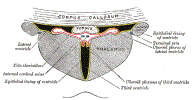
Choroid Plexus
- Produces cerebrospinal fluid in the walls of the Cerebral Ventricles
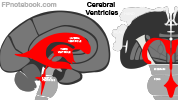
III. Anatomy: Cerebrospinal spinal fluid pathway
- Images
-
General
- CSF is produced in the Choroid plexus of the two Lateral Ventricles
- CSF flows from Lateral Ventricles via foramina to the central Third Ventricle
- From the central Third Ventricle to the Aqueduct of Sylvius and then the Fourth Ventricle
- CSF flows into the subarachnoid space and into the Cerebral Sinuses
- CSF returns to the venous system via reabsorption at the arachnoid villi (esp. in Superior Sagittal Sinus)
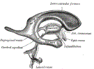
- Lateral Ventricles (left and right)
- Most superior ventricle
- Cerebrospinal fluid originates here (and in the third and Fourth Ventricles)
- Drains via the interventricular foramina (foramina of monro) into the Third Ventricle
- Third Ventricle (midline)
- Drains via the Aqueduct of Sylvius into the Fourth Ventricle
- Fourth Ventricle (midline, posterio fossa)
- Drains out of the intracranial cavity into the subarachnoid space via three openings
- Middle Foramen of Magendie (midline) openings
- Lateral Foramena of Luschka (right and left) openings
- Subarachnoid Space
- CSF flows into the subarachnoid space
- CSF drains into the Superior Sagittal Sinus via arachnoid villi or granules (small holes in the sinus wall)
- Subarachnoid Cistern
- Wider pockets of subarachnoid space and cerebrospinal fluid
- Largest cistern is in the lumbar region (below the L2 Vertebrae)
- Allows for safer Lumbar Puncture

IV. Pathophysiology
-
Hydrocephalus
- Occurs when CSF flow is obstructed, leading to expansion of the Lateral Ventricles
-
Subarachnoid Hemorrhage
- Ruptured Cerebral Aneurysm results in CSF Red Blood Cells
- With time, yellowing develops of the spinal fluid (Xanthochromia)
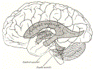
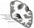
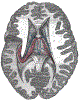
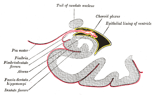
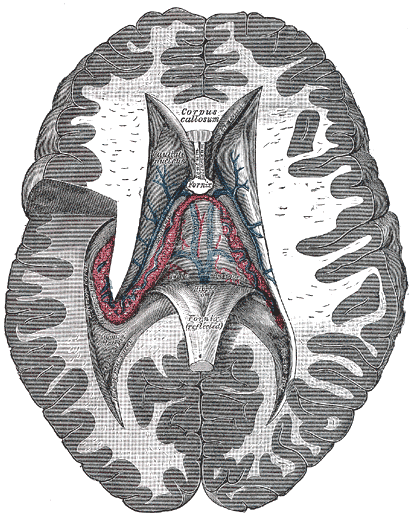
V. Anatomy: Ventricles
-
-
 Lewis (1918) Gray's Anatomy 20th ed (in public domain at Yahoo or BartleBy)
-
 Lewis (1918) Gray's Anatomy 20th ed (in public domain at Yahoo or BartleBy)
-
 Lewis (1918) Gray's Anatomy 20th ed (in public domain at Yahoo or BartleBy)
-
 Lewis (1918) Gray's Anatomy 20th ed (in public domain at Yahoo or BartleBy)
-
 Lewis (1918) Gray's Anatomy 20th ed (in public domain at Yahoo or BartleBy)
-
 Lewis (1918) Gray's Anatomy 20th ed (in public domain at Yahoo or BartleBy)
-
 Lewis (1918) Gray's Anatomy 20th ed (in public domain at Yahoo or BartleBy)


VI. References
- Gilman (1989) Manter and Gatz Essentials of Neuroanatomy and Neurophysiology, Davis, p. 232-7
- Goldberg (2014) Clinical Neuroanatomy, Medmaster, p. 6-15
- Netter (1997) Atlas Human Anatomy, ICON Learning, p. 102-3