
II. Images
-
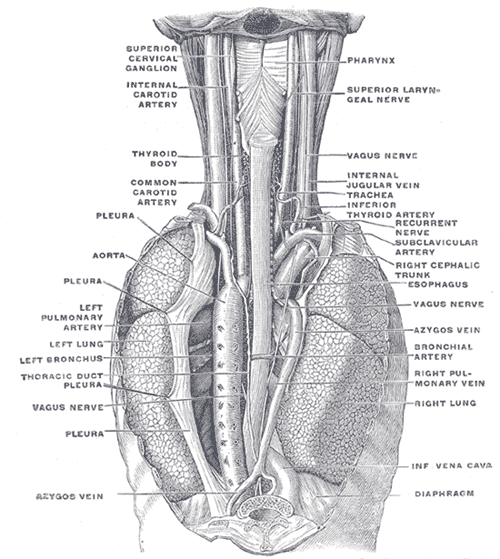
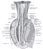 Lewis (1918) Gray's Anatomy 20th ed (in public domain at Yahoo or BartleBy)
III. Anatomy
- Esophagus is divided into three regions
- Cervical Esophagus
- Thoracic Esophagus
- Abdominal Esophaus
- Esophagus wall has three layers
- Mucosa (inner most layer)
- Submucosa
- Muscle
- Striated Muscle in the upper third of Esophagus
- Striated and Smooth Muscle in the middle third of Esophagus
- Smooth Muscle in the lower third of the Esophagus
- Innervation
- Sympathetic innervation (spinal nerves)
- Cervical Esophagus
- Parasympathetic innervation (Vagus Nerve)
- Thoracic and abdominal Esophagus
- Sympathetic innervation (spinal nerves)
- Arterial Perfusion
- Inferior Thyroid artery supplies the upper Esophagus (proximal)
- Aorta supplies the middle Esophagus
- Left gastric artery supplies the lower third of the Esophagus
IV. Pathophysiology
- Food Impaction Sites
-
Diverticular Sites
- Upper Esophageal Sphincter
- Zenker Diverticulum
- Upper Esophagus below circular esophageal Muscle
- Laimer Diverticulum
- Lower Esophageal Sphincter
- Epiphrenic Diverticulum
- Upper Esophageal Sphincter
V. References
- Hagen and Pickle (2023) Crit Dec Emerg Med 37(6): 24-9